Preexcitation Pattern
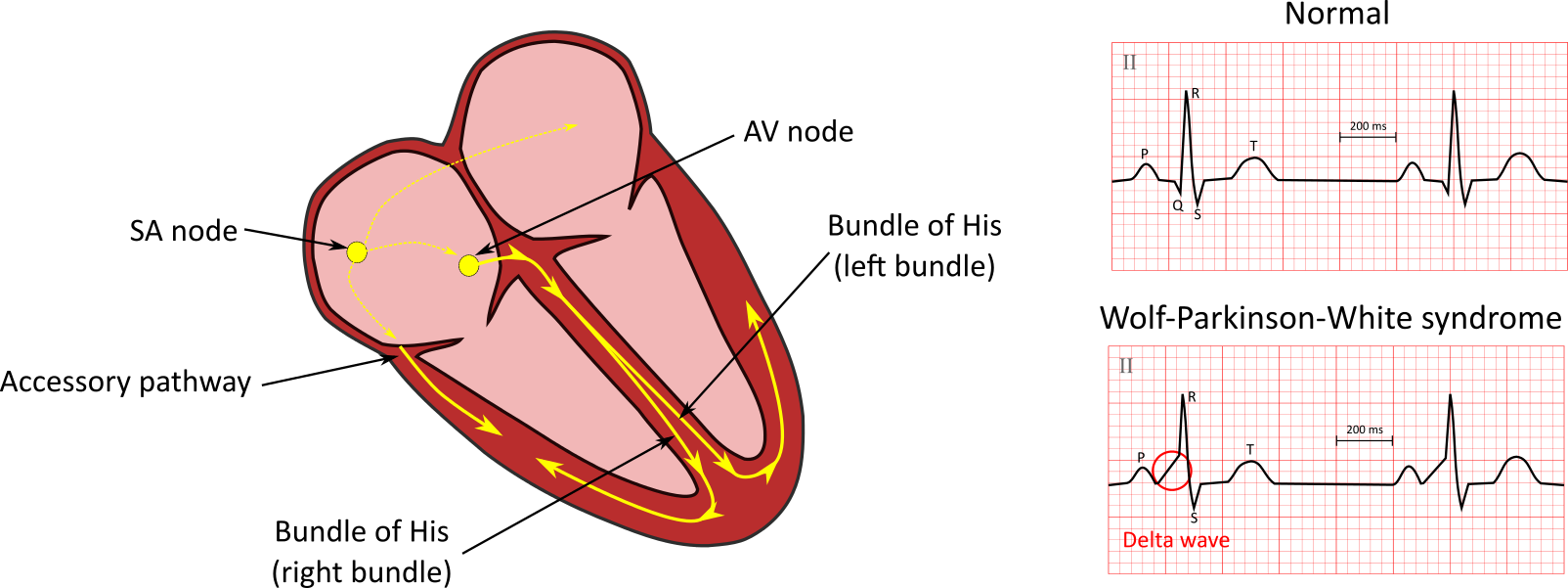
The PR interval in WPW syndrome is short, i.e., < 0.12 seconds. The ECG displays a delta wave pattern with a prolonged distorted QRS complex. Patients also present with alterations in repolarization.
The term preexcitation refers to ventricular activation by an atrial impulse that is conducted through the accessory pathway. The impulse bypasses the AV node. The impulse travels through the accessory pathway faster than the regular impulse passes through the AV node. Consequently, the PR interval is short.
With this abnormal excitation of the ventricles, the QRS complex is distorted because there are alterations in repolarization. The early and direct impulses that cause myocardial activation become fused. The direct impulse comes from the AV node. The early impulse comes from the accessory pathway and slows down the initial phase of ventricular activation.
The upstroke of the QRS complex is slurred due to the slow muscle-fiber-to-muscle-fiber conduction. This is also the cause of delta wave formation. If the accessory pathway impulse conducts more quickly than the normal impulse, then more of the myocardium will depolarize. This causes a wider, more prominent delta wave and a longer QRS complex.

Wolff-Parkinson-White Syndrome 39
Some accessory pathways do not conduct impulses in the antegrade direction but in the retrograde direction (from the ventricle into the atrium) instead. When retrograde conduction is present, the classic WPW pattern is not observed. This is known as a concealed accessory pathway, which is diagnosed using electrophysiologic testing, ventricular ectopy, or ventricular pacing.
Preexcitation and delta waves may not be apparent in some patients with WPW syndrome while in sinus rhythm. These patients have a left-lateral bypass tract with an antegrade direction of conduction. Preexcitation is minimized since the impulse from the accessory pathway takes longer to reach the ventricles than the impulse from the AV node.
Sometimes, the delta waves represent an atypical conduction pathway that does not connect in the usual atrioventricular fashion. The “Mahaim fiber,” for example, is an atriofascicular pathway found along the right lateral atrioventricular groove. It traverses along the right ventricular free wall, connects to the distal portion of the right bundle-branch, and only conducts in the anterograde direction. It is known to be involved in antidromic tachycardia, sending impulses through the normal conductive tissues as part of the retrograde pathway of the circuit.
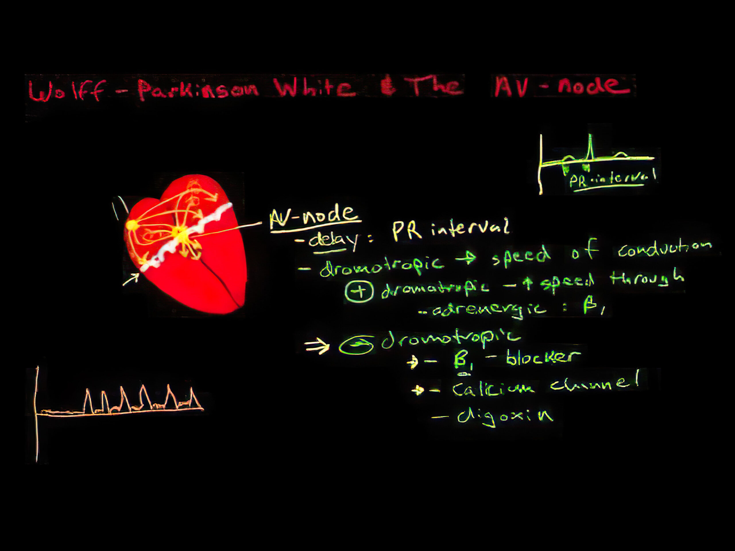
Exercise rarely causes an abrupt loss of preexcitation when the heart rate increases. However, this does not mean that the accessory pathway is unlikely to produce atrial fibrillation with a significant rapid ventricular response. Moreover, during exercise, sympathetic tone is increased, and vagal tone is decreased. These changes in tone influence AV nodal conduction.

Pathophysiology of Wolff-Parkinson-White Syndrome 40
39 Atrioventricular reentry tachycardia. Natalie’s casebook website. Accessed May 8, 2021.
http://www.nataliescasebook.com/tag/atrioventricular-reentry-tachycardia
40 Wolff A. Pathophysiology of Wolff-Parkinson-White syndrome. YouTube website. Accessed September 24, 2020.