ECG Criteria for Biventricular Hypertrophy
ECG Criteria for Biventricular Hypertrophy
When evaluating the 12-lead for signs of biventricular hypertrophy, the clinician looks for:8
Criteria 1: Sokolow-Lyon voltage:9 SV1 + RV5(or RV6) > 35 mm combined with a QRS right axis deviation in patients older than 30 years of age.
Criteria 2: SV6 ≥ 7 mm (without RBBB); this finding can also present in patients with isolated RVH
Criteria 3: Presence of RVH patterns + left atrial enlargement with a P duration of ≥ 0.12 seconds
- S/R ≥ 1 in V5/V6 + LA enlargement
- SV6 ≥ 7 mm + LA enlargement
- ÅQRSF > +90° + LA enlargement (if RBBB is present, then the ÅQRSF
- is calculated on the first 0.06 seconds of the QRS)
This criterion has a specificity of 80% with extremely low sensitivity. Echocardiography is generally required to confirm a diagnosis.
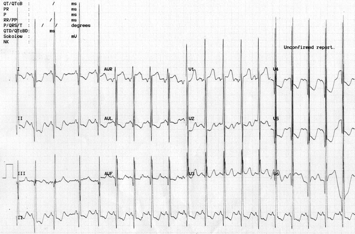
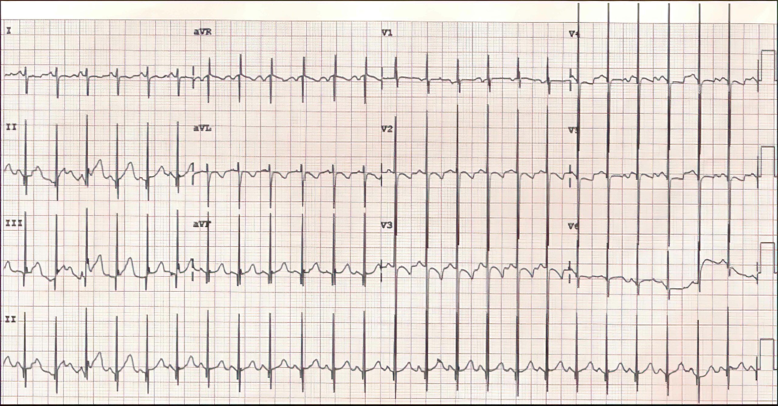
A shallow SV1 and a deep SV2 are additional ECG characteristics of biventricular hypertrophy (see Figure 2). The shallow SV1 is caused by the cancellation of vectors between the extremely large right and left ventricles. This cancellation phenomenon is not apparent in V2 because of its projection, which leads to a deep SV2 instead.
Inadvertent lead placement of V1 can also cause these findings if the lead is connected too high, too low, or too far to the lateral side of the patient.

Biventricular Hypertrophy With Large Biphasic Complexes

12-Lead ECG of a Pediatric Patient Born With a Ventricular Septal Defect
8 Gertsch M. The ECG manual: an evidence-based approach. Springer; 2009.
9 Sokolow M, Lyon TP. The ventricular complex in left ventricular hypertrophy as obtained by unipolar precordial and limb leads. Am Heart J. 1949;37(2):161–186.
https://www.sciencedirect.com/science/article/abs/pii/0002870349905621