Ebstein Anomaly
Ebstein Anomaly
The ECG of a patient with Ebstein anomaly resembles extreme “p pulmonale.” Tall P waves appear in leads III and aVF. Tall, peaked P waves appear in leads V1 and V2. The QRS complexes of leads III and/or aVF have an “M” configuration. Right axis deviation is usually present, although left axis deviation may be present instead.

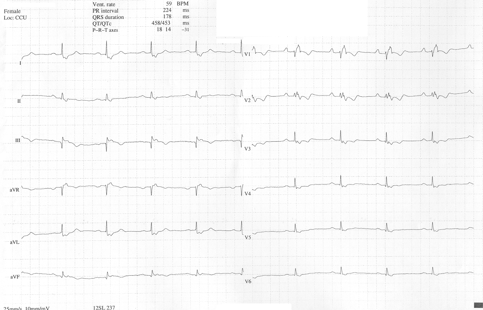
Ebstein Anomaly ECG 64
The ECG in Figure 4 is from a patient with Ebstein anomaly. Signs of right atrial enlargement are evident. Tall P waves (> 2.5 mm) are seen in limb leads II, III, and aVF. First-degree AV block and RBBB are present. T wave inversion appears in precordial leads V1 through V4 along with marked Q waves in lead III, all characteristic of Ebstein anomaly.
64 Gibson CM. Ebstein’s anomaly of the tricuspid valve electrocardiogram. Wikidoc website. Accessed September 30, 2020.