Differential Diagnosis of Wolff-Parkinson-White Syndrome
Myocardial Infarction
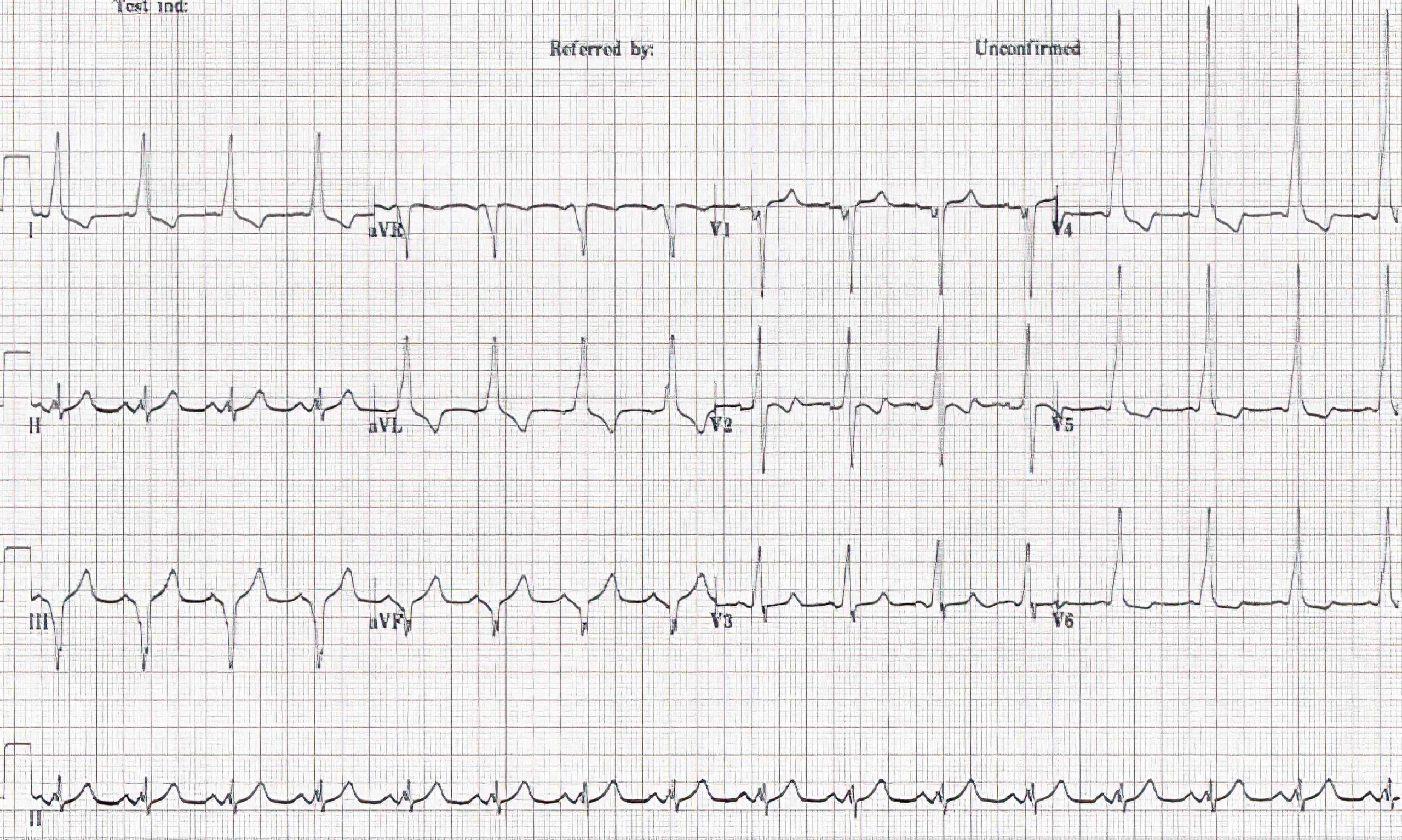
Patients with WPW syndrome often have ECGs with pathologic Q waves that may be mistaken for chronic MI. The presence of the classic signs of WPW syndrome should guide the clinician’s diagnosis. WPW syndrome presents with positive T waves in the leads where there are pathologic Q waves. In patients with chronic myocardial infarction, the T waves have a negative deflection.
Frontal leads III and aVF have a downward QRS complex with a negative delta wave. This can also be mistaken for chronic inferior wall MI (see ECG in figure 4 below). Tall R waves in precordial leads V1 through V3 may be mistaken for posterior, inferoposterior, and posterolateral wall MI.

Type B WPW Syndrome ECG 44
Chronic MI is difficult to ascertain on ECG when present in patients with preexcitation. On the other hand, ST elevation in acute MI is easily detected.
Left Ventricular Hypertrophy
Preexcitation in WPW syndrome can produce tall R waves on ECG. This can be mistaken for LVH (see the ECG in figure 2 above). Asymmetric T waves may also cause the clinician to question whether there is a strain pattern.
Pseudo-Delta Wave
Pseudo-delta waves are not due to preexcitation in the presence of delta waves in precordial leads V2 and V3, and frontal leads II, III, and aVF. These waves are due to the “projection” pattern. This normal variant can be differentiated from WPWsyndrome if the PR interval and QRS complex are normal.
44 Burns E. Preexcitation syndromes. Life in the fastlane website. Accessed September 24, 2020.
https://litfl.com/wp-content/uploads/2018/08/WPW-Type-B-ECG-2.jpg