Hypokalemia
Hypokalemia is a common clinical problem. Potassium enters the body through oral intake or intravenous infusion. It is stored in the cells and excreted in the urine. Decreased intake, increased translocation of potassium into cells, and potassium loss in the urine, gastrointestinal tract, or sweat lower the serum potassium concentration. Severe dehydration from diarrhea or vomiting is a common cause of hypokalemia. Patients treated with diuretics may also present with hypokalemia.
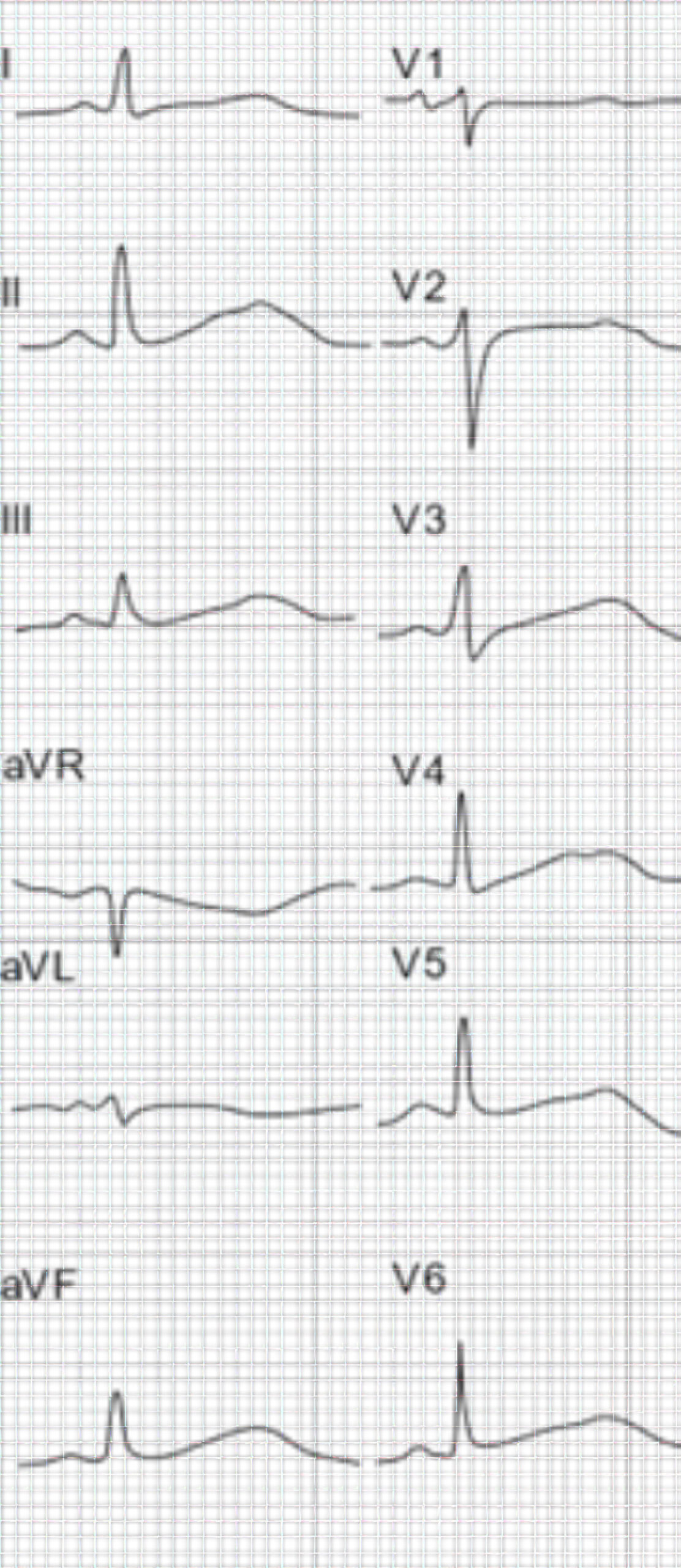
T wave fusion and a prominent U wave are ECG changes associated with hypokalemia. Since the end of the T wave is not delimited, the QT interval is immeasurable. ST depression may be visible at times.
Other causes of TU fusion include congenital long QT syndrome and polymorphic ventricular tachycardia. If the patient with polymorphic ventricular tachycardia does not revert to normal sinus rhythm, the patient may develop syncope or progress to ventricular fibrillation. Patients on amiodarone may also exhibit ECG changes resembling hypokalemia.

ECG depicts complete fusion of the TU waves.
Case 3

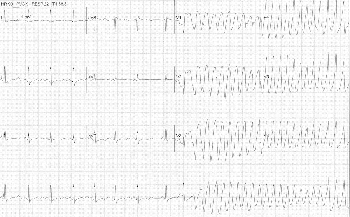
Severe Hypokalemia ECG Rhythms. 30
The ECG above was obtained from a patient with severe hypokalemia (K+ = 1.7 mmol/L). The ECG shows inverted T waves, prominent U waves, and a long Q-U interval. A PAC has landed on the end of the T wave and may be the cause of the R-on T phenomenon. R-on-T, in turn, instigated the polymorphic ventricular tachycardia. This ECG can be classified as torsades de pointes.
Severe hypokalemia is treated with oral potassium and magnesium. A temporary pacemaker may be needed until the patient recovers, and the underlying cause of hypokalemia is treated.
30 Burns E, Buttner R. Polymorphic VT and torsades de pointes. Life in the fastlane website. Accessed August 22, 2020.
https://litfl.com/wp-content/uploads/2018/08/ECG-hypokalaemia-torsades-2.jpg