Changes in the T Wave
Negative T Waves
Asymmetric, negatively deflected T waves that appear in most of the limb leads are nonspecific and usually normal.
- Negative T waves are a normal finding in limb leads II, III, aVF, and aVL.
- Inverted T waves are also a normal finding V.
- Inverted T waves in V2 and V3 are a normal finding in healthy men ≤ 25 years old and in healthy women ≤ 35 years old.
Negative T waves in limb lead I may be caused by cardiac disease. Negative T waves in leads V2 and V3 may be caused by a right ventricular pathology, such as an atrial septal defect, pulmonary embolism, or an arrhythmogenic right ventricle.

76-Year-Old Man With CAD and COPD ECG

63-Year-Old Woman With Hypertension and Aortic Stenosis ECG
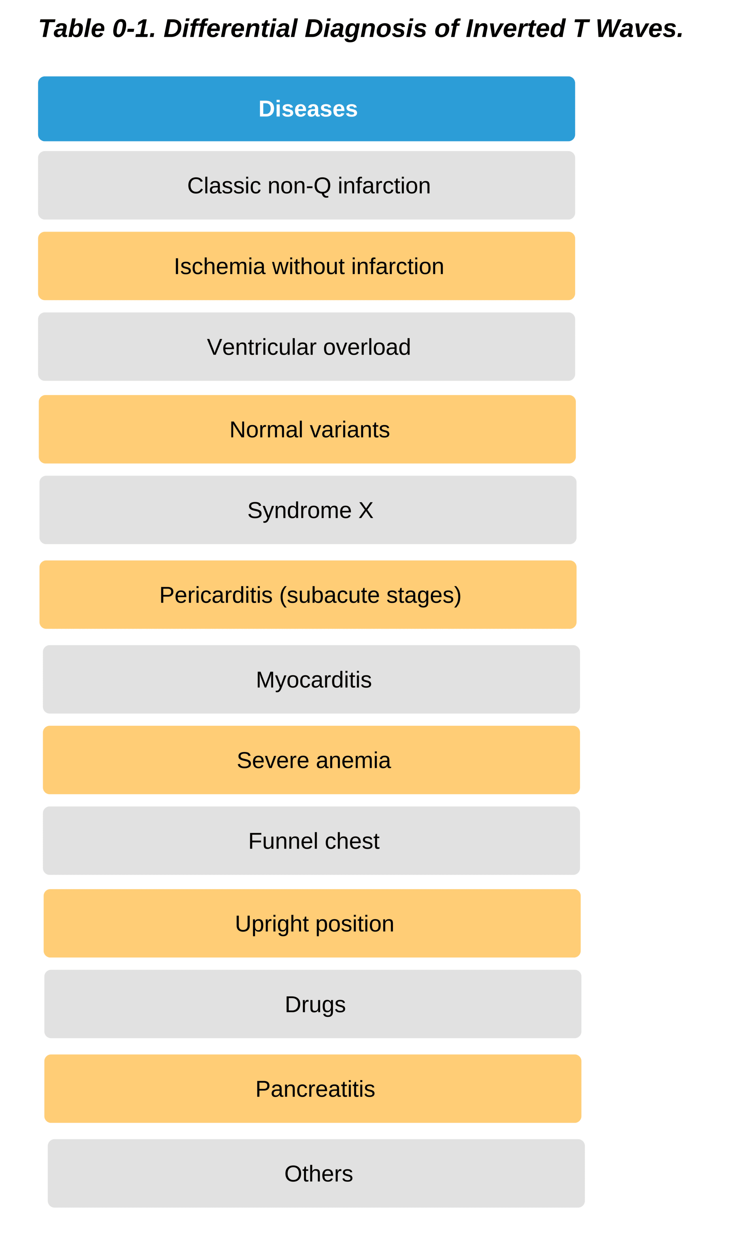
The term primary negative T waves is used to describe ECG findings associated with ischemia. The term secondary negative T waves is used when describing ECG findings suggestive of ventricular hypertrophy. In patients with myocardial ischemia, the inverted T waves are usually symmetrical. In patients with LVH or left ventricular overload, the inverted T waves are usually asymmetrical. But this is not always the case.
Symmetrical inverted T waves have been documented in patients with subacute pericarditis and in those with hypertrophic cardiomyopathy. Asymmetrical inverted T waves have been noted in patients with an assortment of illnesses and inflammations. They are also associated with the effects of certain drugs and can be seen in patients with normal hearts.
Elevated T Waves
Hyperkalemia is the most common cause of symmetrical, abnormally elevated T waves. They are also seen in the early stages of STEMI. After the onset of symptoms, these elevated T waves last only a few minutes before significant ST elevations evolve.
Elevated T waves in V2 and V3 are usually normal variants and are often seen in patients with sinus bradycardia. In the case below, a patient was diagnosed with Prinzmetal or vasospastic angina.
Prinzmetal angina is characterized by chest pain at rest and attributed to sudden coronary artery vasospasm, which is usually relieved by sublingual nitrates. If the ECG of a patient with Prinzmetal angina can be obtained before the onset of ST segment elevation, tall peaked T waves may be noted as in the case below.

Hyperacute T waves are present in a patient with Prinzmetal or vasospastic angina. 28
Giant Negative T Waves
A negative T wave with an amplitude of 5–15 mm is a rare occurrence. Such giant negative T waves are found in patients with LVH brought about by hypertension or hypertrophic cardiomyopathy and in patients with apical hypertrophy.
Giant negative T waves without any other concomitant ECG abnormalities have been documented in patients with non-Q wave infarction patterns. They may be brought about by pacemakers (Chatterjee phenomenon) and also may appear in some patients with cerebral diseases and pulmonary edema.
Global T wave inversion in women is of no significant consequence. Studies have postulated that it is catecholamine-related and has a good prognosis. 29
Discrepancies in QT Duration
Giant T wave inversions with excessive QT prolongation are rare. This ECG pattern is closely related to postsyncopal bradycardia syndrome. It can manifest in patients with subarachnoid hemorrhage, trauma, iatrogenic injuries, and during epileptic episodes.
If the QT interval is too long or too short, there will be changes in repolarization patterns. The ST segment may be isoelectric, elevated, or depressed if a patient has congenital long QT syndrome or acquired long QT syndrome. Acquired long QT syndrome is a result of hypokalemia and a side effect of antiarrhythmic cardiac medications. Congenital short QT syndrome is a very rare condition.
28 Burns E. T wave. Life in the fastlane website Accessed August 24, 2020.
https://litfl.com/wp-content/uploads/2018/08/Hyperacute-T-waves-due-to-anterior-STEMI.jpg
29 Walder LA, Spodick DH. Global T wave inversion. J Am Coll Cardiol. 1991;17(7):1479–1485.