Chapter Progress
0% Complete
Get 12-Lead ECG Certified Today
Trifascicular Block (RBBB + LAFB + LPFB)
Trifascicular block is a rare occurrence. Survival is maintained if an atypical medial fascicle is present. The medial fascicle is located between the left anterior and posterior fascicles. Patients may develop complete AV block at any time.
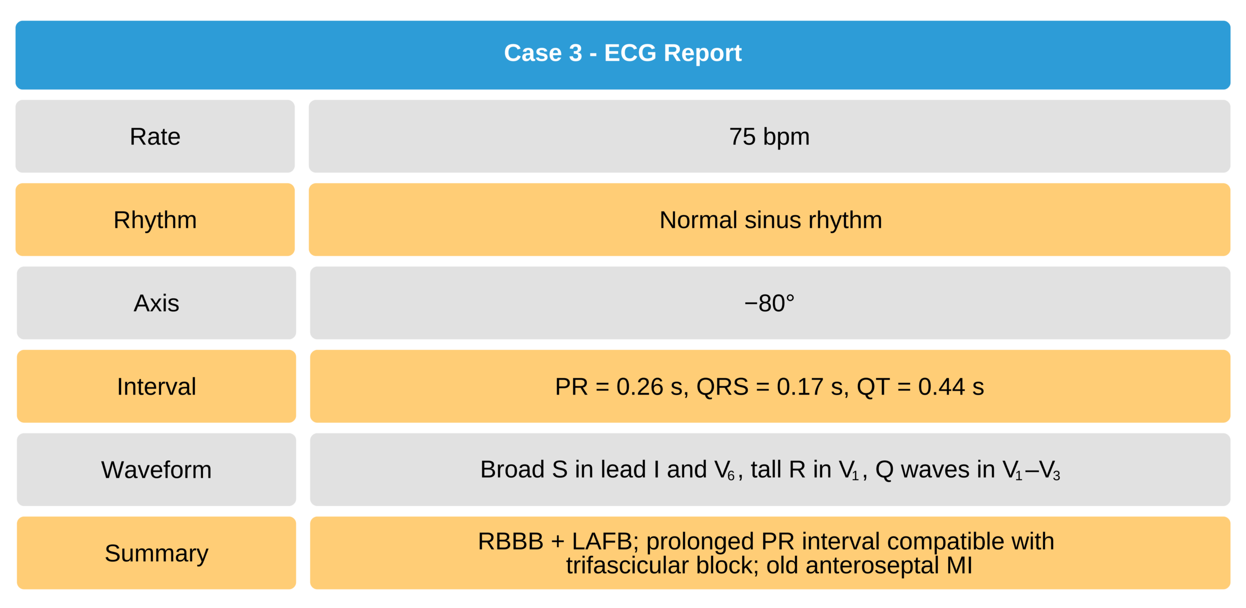
Trifascicular block is difficult to diagnose. The ECG findings show a bilateral bifascicular block with a prolonged PR interval. More invasive electrophysiological studies must be performed to distinguish trifascicular block from bilateral bifascicular block.
ECG Findings
- rSr’ or rsR’ in V1
- Frontal QRS axis depicting left axis deviation (−30° to −90°)
- Slurred R downstroke in leads I, aVL, V5, and V6 following an s wave of reduced duration
- Prolonged PR interval
- PQ interval not prolonged

Trifascicular Block
Case 3

Trifascicular Block ECG Rhythms Case 3