Chapter 1: Theories in ECG Interpretation
6 Topics | 1 Quiz
Chapter 2: Systematic Approach to ECG Interpretation
2 Topics | 1 Quiz
Chapter 5: Abnormalities of the P Wave
6 Topics | 1 Quiz
Chapter 6: Left Ventricular Hypertrophy
3 Topics | 1 Quiz
Chapter 8: Biventricular Hypertrophy
4 Topics | 1 Quiz
Chapter 9: Acute Pulmonary Embolism
7 Topics | 1 Quiz
Chapter 10: Fascicular Blocks
7 Topics | 1 Quiz
Chapter 11: Complete and Incomplete Bundle-Branch Blocks
5 Topics | 1 Quiz
Chapter 14: Myocardial Infarction
10 Topics | 1 Quiz
Chapter 16: Pericarditis
6 Topics
Chapter 19: Atrial Arrhythmias
5 Topics | 1 Quiz
Chapter 20: Sick Sinus Syndrome
10 Topics | 1 Quiz
Chapter 22: Atrioventricular Junctional Tachycardias
5 Topics | 1 Quiz
Chapter 23: Premature Ventricular Contractions
7 Topics | 1 Quiz
Chapter 24: Ventricular Tachycardia
3 Topics
Chapter Progress
0% Complete
Get 12-Lead ECG Certified Today
Right Bundle-Branch Block with Left Anterior Fascicular Block
ECG Findings
- QRS duration > 0.12 seconds
- rsR’ complex in V1 (as seen in RBBB)
- Sometimes a slurred R wave or a qR complex in V1
- Sometimes the S wave in V6 is a mirror image of that in lead V1
- Left axis deviation
- Small q waves in aVL and rS complex in leads II and III
- Positive T waves in aVF
- Clockwise rotation in the precordial leads, commonly with an rS complex absent Q wave in leads V5 and V6

Bilateral Bifascicular Block of the RBBB + LAFB Type
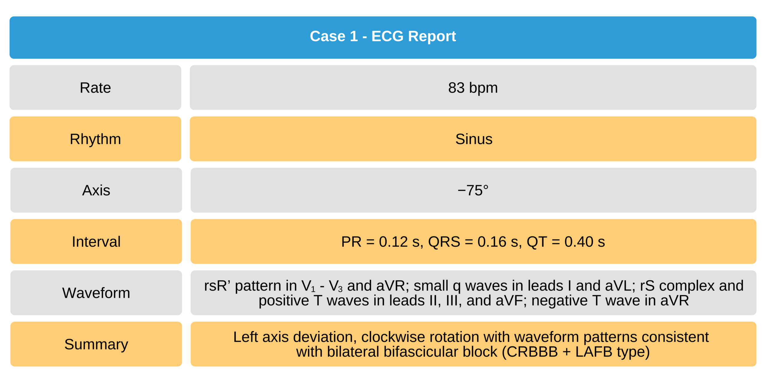
Case 1
67-year-old man with bilateral bifascicular block (complete RBBB + LAFB type)

67-Year-Old Man With Bilateral Bifascicular Block ECG
Figure 2.
Table 0-1 ECG findings of Case 1 (see Figure 2).