Chapter 1: Theories in ECG Interpretation
6 Topics | 1 Quiz
Chapter 2: Systematic Approach to ECG Interpretation
2 Topics | 1 Quiz
Chapter 5: Abnormalities of the P Wave
6 Topics | 1 Quiz
Chapter 6: Left Ventricular Hypertrophy
3 Topics | 1 Quiz
Chapter 8: Biventricular Hypertrophy
4 Topics | 1 Quiz
Chapter 9: Acute Pulmonary Embolism
7 Topics | 1 Quiz
Chapter 10: Fascicular Blocks
7 Topics | 1 Quiz
Chapter 11: Complete and Incomplete Bundle-Branch Blocks
5 Topics | 1 Quiz
Chapter 14: Myocardial Infarction
10 Topics | 1 Quiz
Chapter 16: Pericarditis
6 Topics
Chapter 19: Atrial Arrhythmias
5 Topics | 1 Quiz
Chapter 20: Sick Sinus Syndrome
10 Topics | 1 Quiz
Chapter 22: Atrioventricular Junctional Tachycardias
5 Topics | 1 Quiz
Chapter 23: Premature Ventricular Contractions
7 Topics | 1 Quiz
Chapter 24: Ventricular Tachycardia
3 Topics
Chapter Progress
0% Complete
Get 12-Lead ECG Certified Today
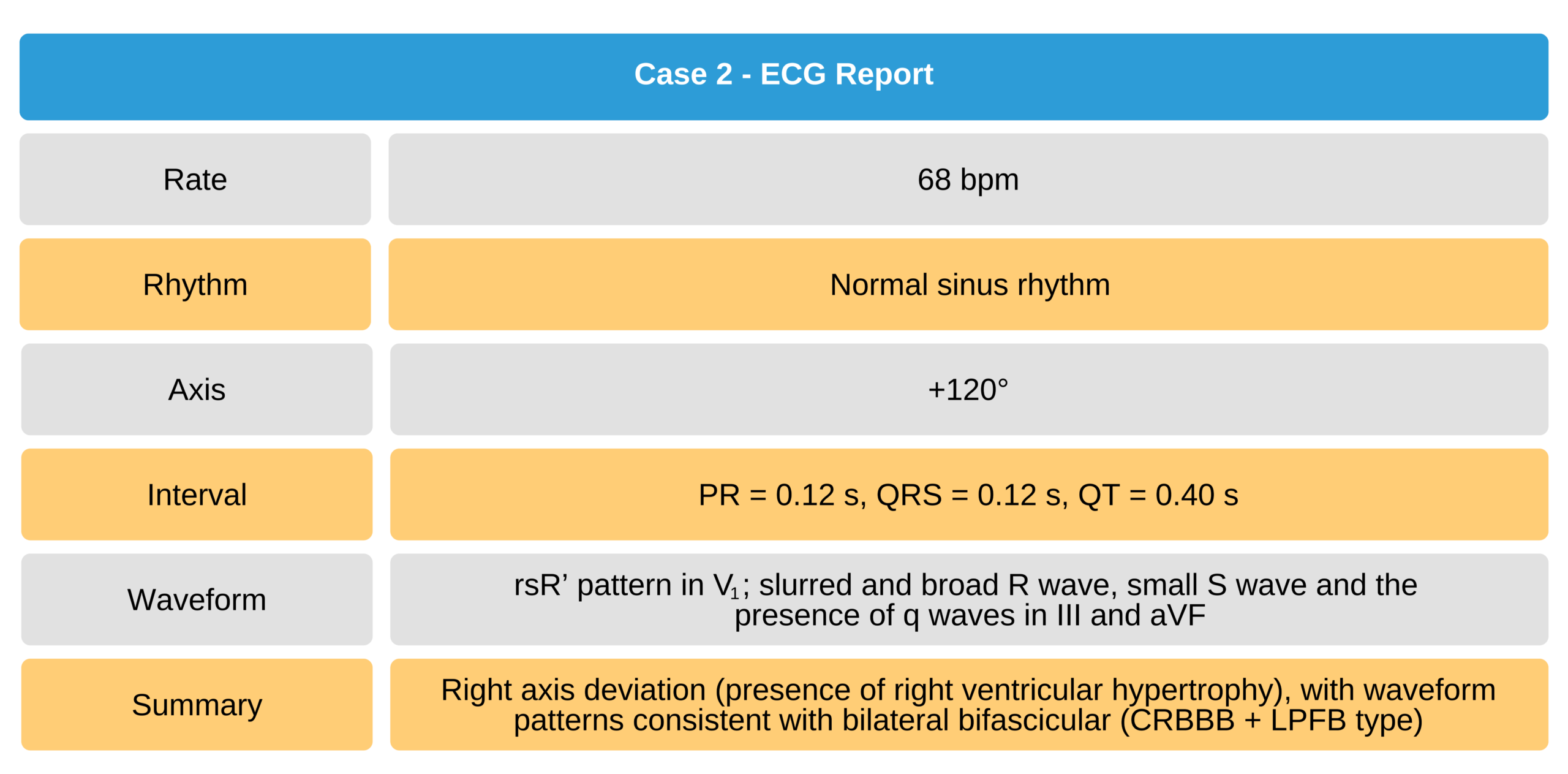
Right Bundle-Branch Block + Left Posterior Fascicular Block
ECG Findings
- Lead V1 has typical RBBB pattern
- Slurred R wave seen in leads III, aVF, V5, and V6 due to delayed activation of the inferior left ventricle
- S wave smaller than normal
- R wave broader than normal
- Q wave preserved
- Inferior wall MI masked by left posterior fascicular block

Bilateral Bifascicular Block of the RBBB + LPFB Type
Case 2
63-year-old man with bilateral bifascicular block (RBBB + LPFB type)

63-Year-Old Man With Bilateral Bifascicular Block ECG