Left Posterior Fascicular Block
Left posterior fascicular block is difficult to diagnose because there are minimal ECG changes. Minute changes in the frontal QRS axis of the limb leads may be mistaken for normal variants. Cardiac diseases, such as inferior wall infarction, are closely associated with left posterior fascicular block with a frontal axis between +50° and +80° and even up to +140°.
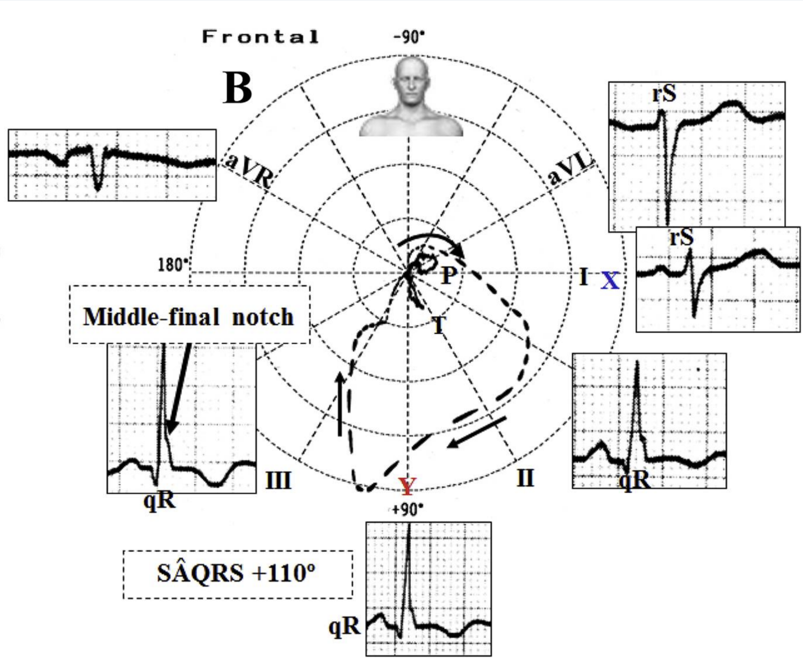
Other ECG findings include a q and T wave of varying duration in leads III and aVF. A slurred R downstroke is seen at times in leads III, aVF, and V6. Lead V6 may have a small or almost absent s wave (see Figure 3).

Left Posterior Fascicular Block With Vectors and Corresponding ECG Tracings 12
A left posterior fascicular block masks inferior wall infarction. It will show tall R waves with absent or slightly enlarged Q waves and negative, flat, or positive T waves in leads II, III, and aVF.
In patients with a left posterior fascicular block but without an inferior wall MI, ECG findings have a pattern of small q waves and greater R waves in the inferior leads. The ECG also has a frontal QRS axis between +80° and +120°.
Clinical conditions depicting a frontal QRS axis between +50° and +80°/+120° may also be confused with left posterior fascicular blocks. This pattern can be seen in a young individual with a small body habitus or in patients with RVH. The correct diagnosis of left posterior fascicular block must coincide with an existing inferior wall MI as described. Other diagnostic imaging modalities may be used to confirm an inferior wall MI, such as myocardial perfusion scintigraphy or 2-D echocardiogram.
12 Pérez-Riera AR, Barbosa-Barros R, Daminello-Raimundo R, de Abreu LC, Tonussi Mendes JE, Nikus K. Left posterior fascicular block, state-of-the-art review: a 2018 update. Indian Pacing Electrophysiol J. 2018;18(6):217–230.