T and ST Wave Changes in Ischemia
Ischemia is a medical condition in which there is poor perfusion for any reason. When an area of the heart is persistently lacking adequate perfusion (e.g., from occluded coronary arteries), the innermost layer of the heart in that area is affected first.
As coronary artery disease (CAD) progresses, an infarct eventually ensues. The infarct affects the middle layer next and finally the superficial layer of the myocardium. If not corrected, the patient will have significant cardiac dysfunction and eventual heart failure.
Figure 6 represents the impact of an infarct on the layers of the cardiac muscle. T and ST wave changes depend on the stage of ischemia and the layers of myocardium that have been impacted.
In other words, T and ST wave changes in ECG tracings help the clinician establish the severity of tissue hypoxia, especially in the left ventricle.
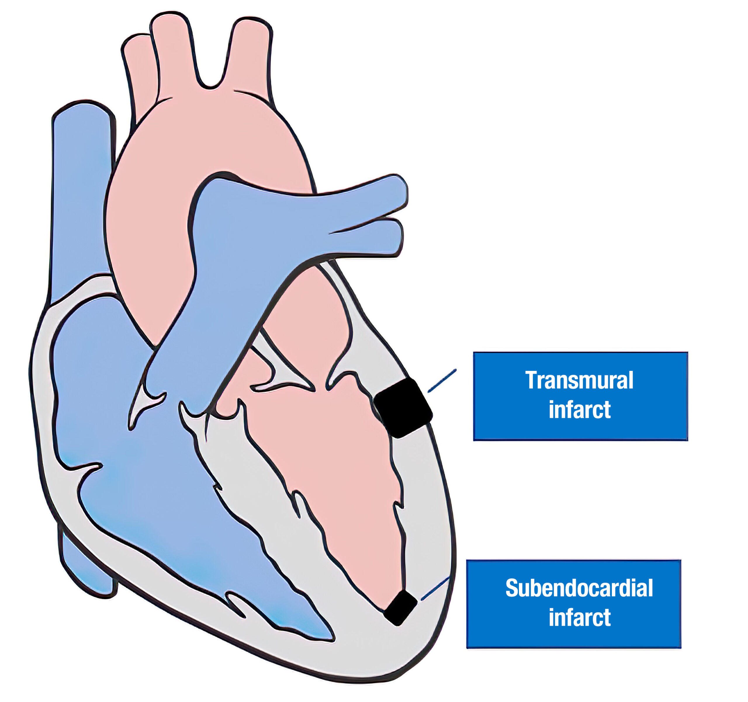
Figure 6 Cardiac Infarct Impact
The Four Degrees of Coronary Artery Disease Insults
Subendocardial ischemia is the lowest grade of ischemia (see Figure 7). The affected area at the subendocardial level is the most superficial layer of the myocardium that can be expressed on ECG. The morphology can be mistaken for moderate hyperkalemia.
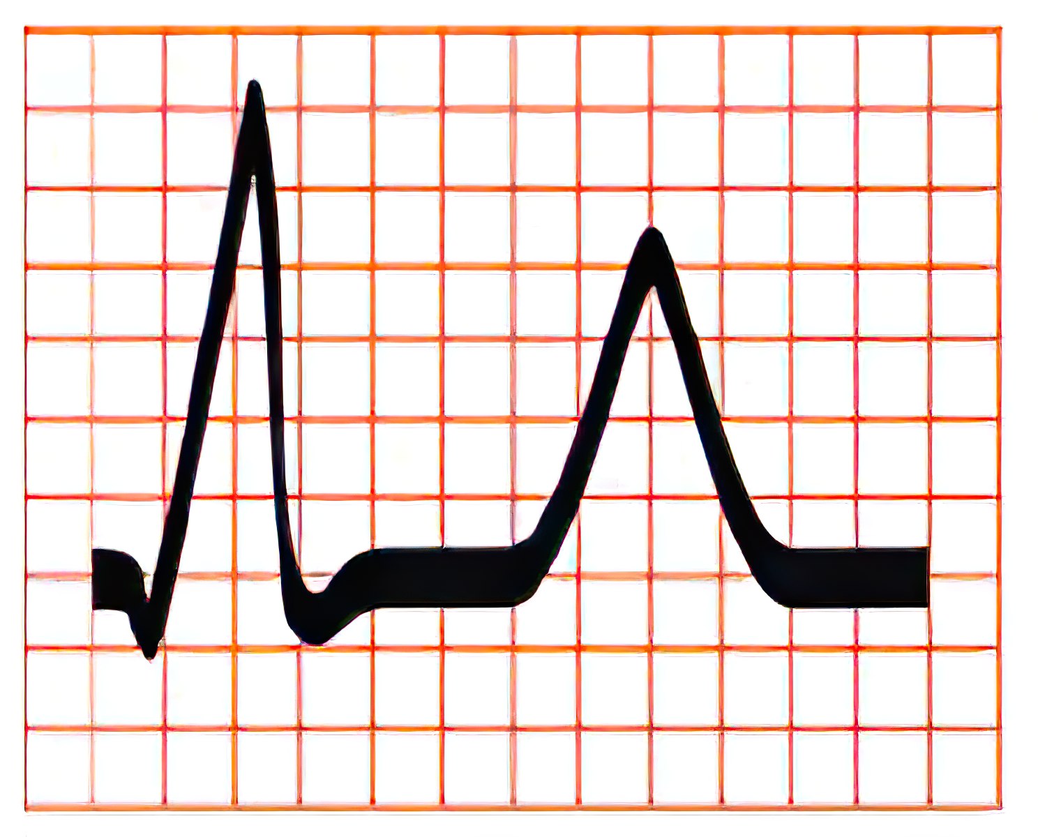
Figure 7 Subendocardial Ischemia ECG
Subepicardial ischemia represents a higher grade of ischemia compared to subendocardial ischemia. ECG morphology shows a pattern of symmetrical inverted T waves (see Figure 8). These specific ECG findings are not exclusive to subepicardial ischemia and may be caused by other cardiac pathology.
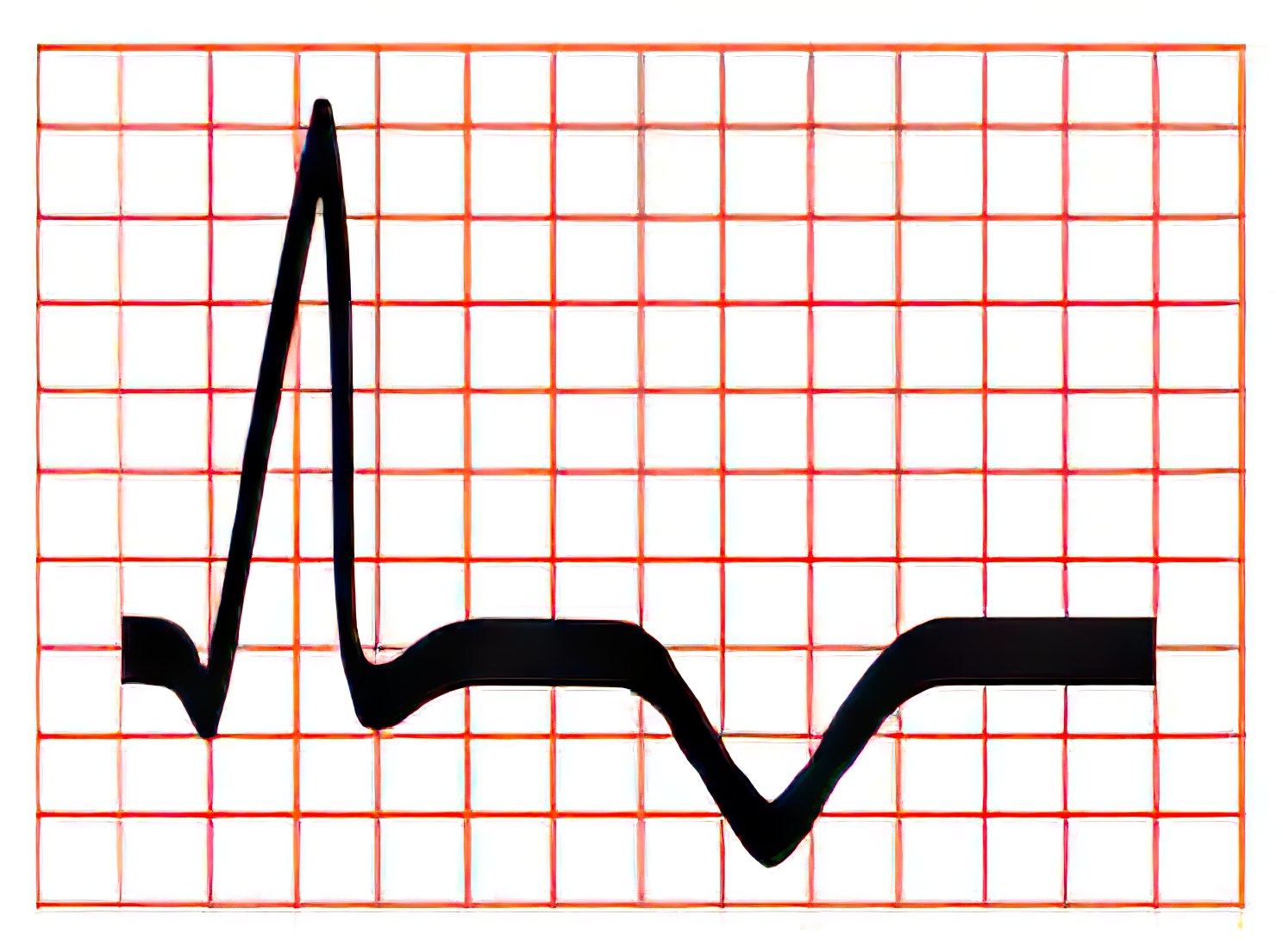
Figure 8 Subepicardial Ischemia ECG
Subendocardial lesion is a higher grade of ischemia compared with subepicardial ischemia. The characteristic ECG pattern of a subendocardial lesion is ST-segment depression (Figure 9). This pattern is also seen in other cardiac conditions such as left ventricular hypertrophy and in patients taking digitalis.
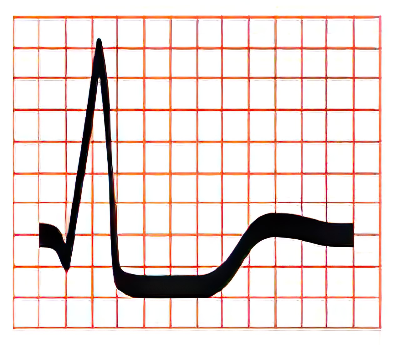
Figure 9 Subendocardial Lesion ECG
Subendocardial ischemia, subepicardial ischemia, and subendocardial lesions are all reversible causes of ischemia when the patient is treated early with fibrinolysis or percutaneous coronary intervention.
Transmural lesion is the highest grade of ischemia and is also known as transmural injury. It encompasses all the layers of the myocardium and is represented by extensive ST segment elevation (see Figure 10).
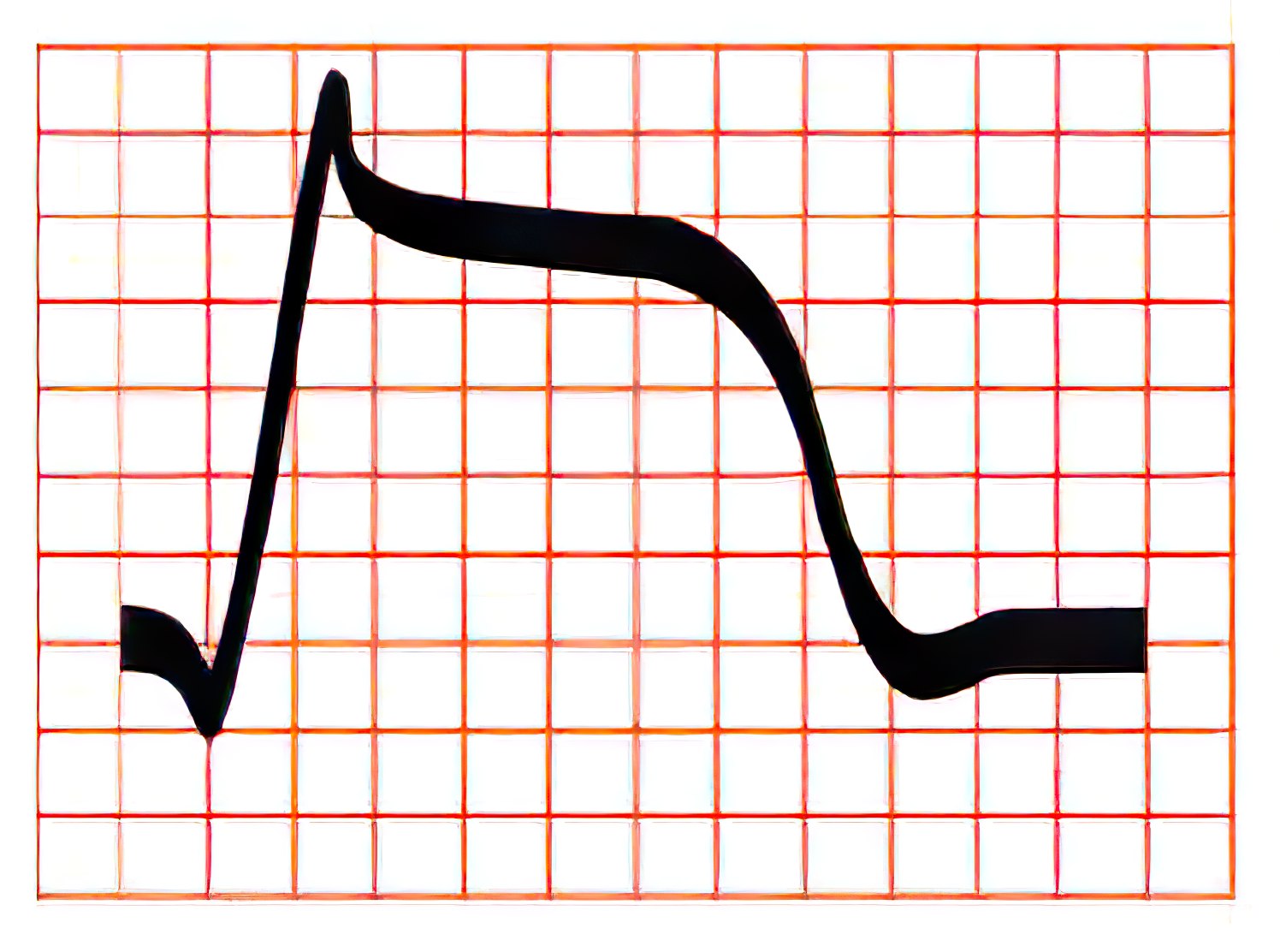
Figure 10 Transmural Injury ST Segment Elevation
This ECG pattern is reversible if the medical condition is caused by vasospasm, also known as Prinzmetal angina.
If this type of ischemia persists, the patient will progress to myocardial infarction (MI). An MI has the appearance of new Q waves in ECG tracings.
ST elevation can also be seen in medical conditions other than coronary artery disease, such as pericarditis and early repolarization.
Lesions involving the left ventricular myocardium have the highest grade electrocardiographic ischemia compared to the rest of the myocardium since anything that disrupts the left ventricular systolic function compromises cardiac output.