Changes in the ST Segment
ST Elevation
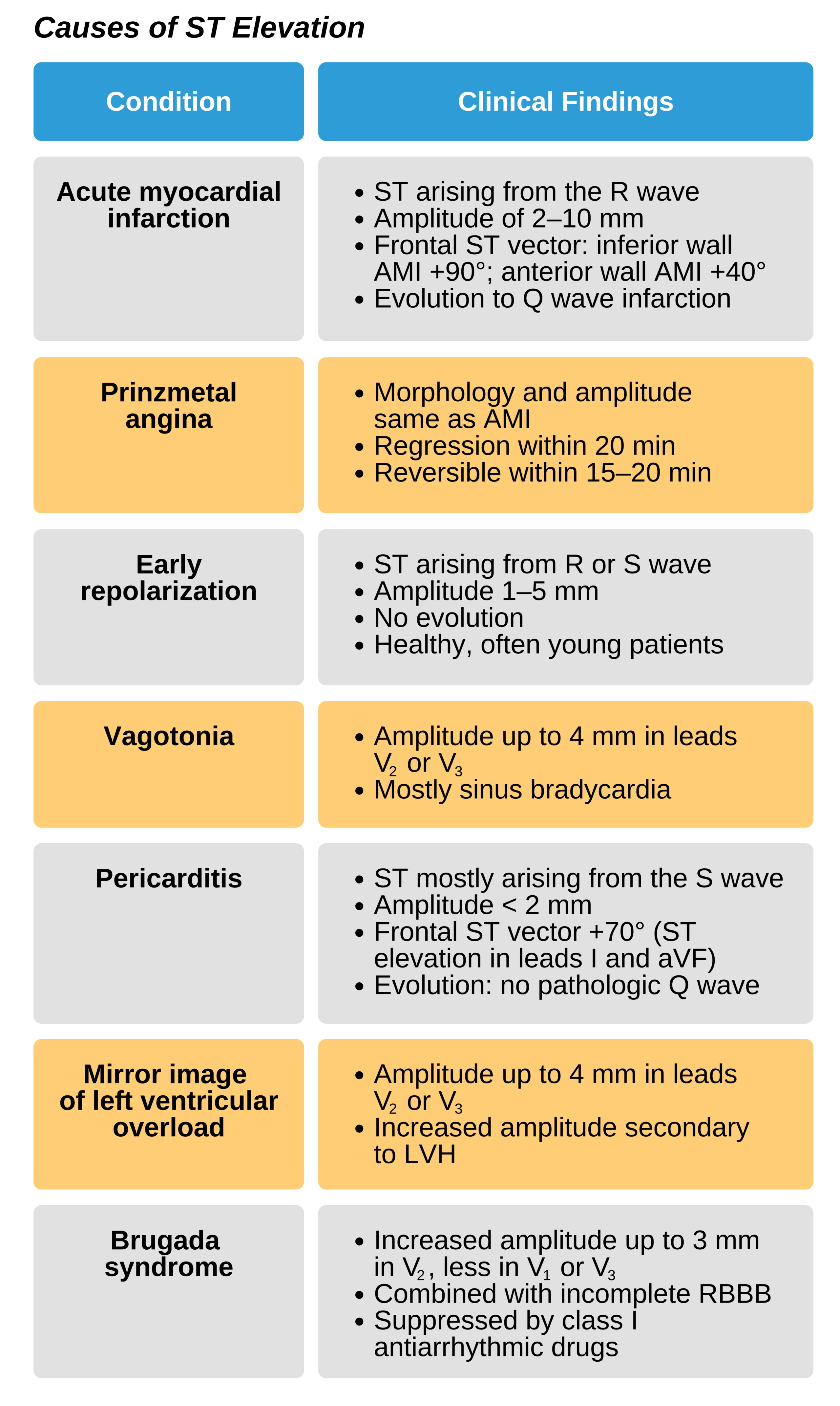
ST segment elevation of 3 mm or more represents acute MI. This finding is also observed in other rare cardiac diseases such as Prinzmetal angina, Brugada syndrome, LVH, and left ventricular overload (lead V2). It can also be found in benign early repolarization. Patients with acute pericarditis may have ST elevation, but the ST segment elevations do not exceed 2 mm.

ST Elevation Within QT Interval
ST Depression
ST depression is an indication of ischemia. When trying to confirm coronary artery disease, clinicians attempt to elicit ST depression during a 12-lead exercise (treadmill) stress test. Anteroseptal wall ischemia cannot be identified on an ECG, but significant ST depression in leads V2 or V3, and sometimes in V1, likely represents a mirror image of impulse activity during an acute posterior wall MI.
ST depression at rest in precordial leads V5 and/or V6 and in limb leads I and/or aVL can be attributed to left ventricular overload with or without LVH. A mirror image in lead V1 and/or V2 may be present.
Digitalis drugs may also elicit a 1 mm ST depression resembling a tub configuration on ECG. In most cases, a 1 mm ST depression is insignificant.