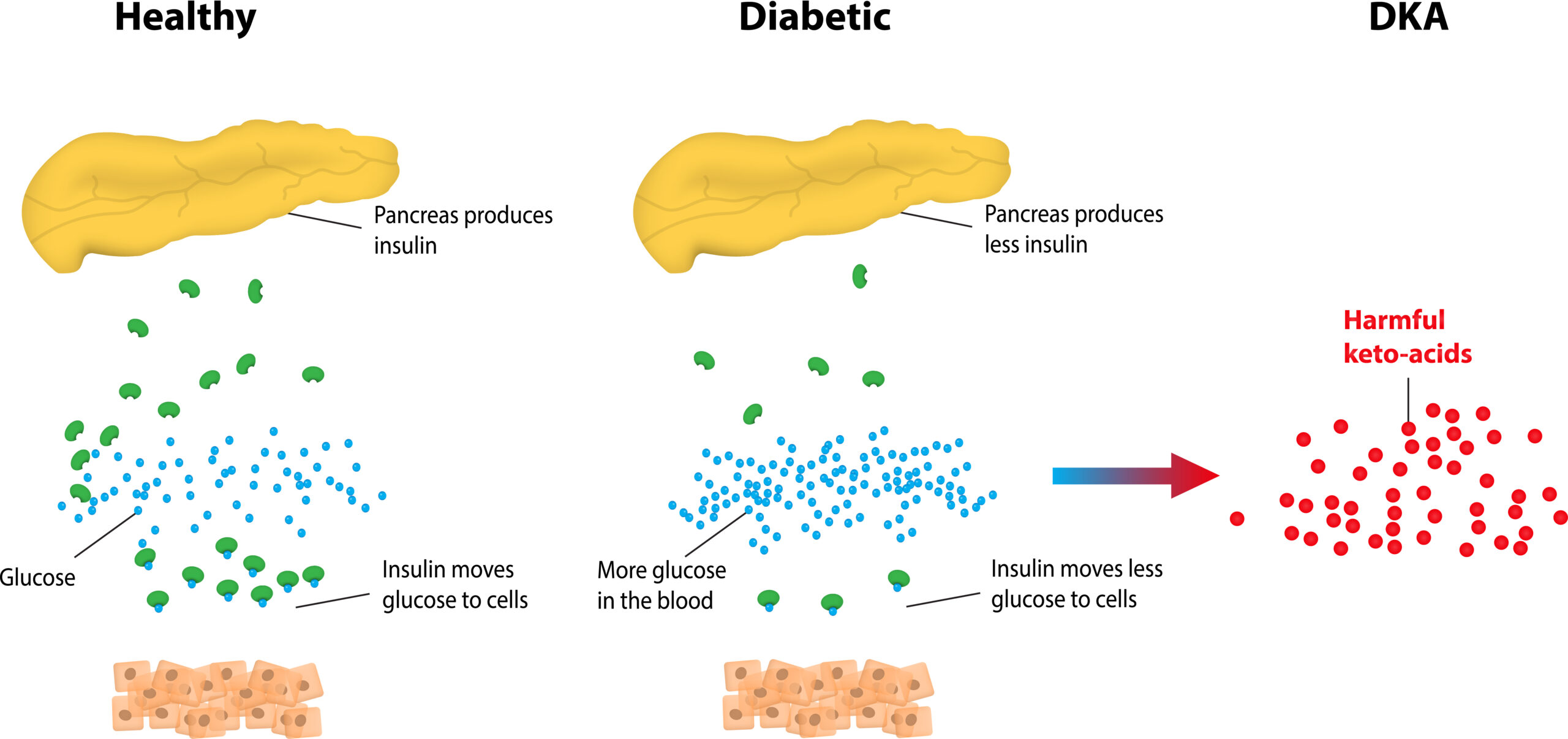
Diabetic Ketoacidosis (DKA)
DKA is a significant diabetes complication that leads to an emergency. DKA is due to very excessive glucose levels and is primarily secondary to the underlying mechanisms of diabetes: insulin insufficiency.
Related Video: What is Diabetic Ketoacidosis (DKA)?
- Hyperglycemia: insulin allows glucose to enter the cell. If there is insufficient or nonfunctional insulin, glucose accumulates outside the cells leading to hyperglycemia. Furthermore, glucagon, which is released due to the lack of insulin, will also stimulate more synthesis of glucose.
- Dehydration: hyperglycemia leads to osmotic diuresis as glucose is excreted into the urine, taking water with it. The patient can have significant dehydration, hypotension, and potentiated acidosis.
- Ketoacidosis: due to insufficient intracellular glucose, the body turns to fats for metabolism. Fats are oxidized into acetoacetic and free fatty acids. Acetoacetic acid converts to ketones, which are acidic. Elevated ketones can lead to ketoacidosis. They also cause the high anion gap of DKA.
- Hypokalemia: total potassium is also lost during osmotic diuresis. There may be normal serum levels as the acidosis of DKA shifts intracellular potassium to the extracellular space. As DKA is treated and acidosis resolves, potassium shifts back intracellularly, which can reveal significant hypokalemia if there is no associated potassium replacement.
Etiologies of DKA
DKA occurs in patients with diabetes. It can be the presentation of the disease as well as can signal a shift from non-insulin-dependent to insulin-dependent diabetes. Another common cause is in a patient who decreases or stops insulin therapy. Some patients will have recurrent DKA suggesting poor control despite optimal medical therapy.
Key Takeaway
Diabetes can initially present as DKA.
Remember that DKA can occur in an undiagnosed diabetic as the presenting sign.
DKA can indicate a shift from non-insulin-dependent to insulin-dependent diabetes.

Diabetic Ketoacidosis Flowchart
Other risk factors or precipitants of DKA include:
- Infection: usually this is a urinary tract infection or pneumonia
- Infarction: Typically, stroke is associated with poor outcomes in insulin-dependent diabetics
- Ignorance: poor compliance or understanding of insulin therapy or dietary recommendation
- Ischemia: acute MI (and associated adrenergic excess) can precipitate DKA
- Intoxication: alcohol abuse can precipitate DKA
- Implantation: pregnancy-associated complications of diabetes are numerous.
Diagnosing DKA
There is a range of DKA symptoms, and consequently, a low threshold for suspicion is needed with insulin-dependent diabetic patients. Patients often will have vague GI complaints, including abdominal pain, nausea, and emesis.
Treating DKA
DKA is life-threatening and should be treated emergently. Initial management should be similar to other emergent conditions
- ABC: ensure an open airway, effective breathing, and adequate circulation. Patients may require advanced airways of ventilation. Dehydration should be managed accordingly.
- Differential: obtain labs, 12 lead EKG, ABGs and urine studies
- Assess vital signs: evaluate for fever, hypotension, arrhythmia, and tachycardia as well as respiratory distress.
- Obtain oxygen saturation and supplement oxygen as needed. Obtain IV access and begin cardiac monitoring. Provide intravascular fluid.
The four abnormalities of DKA discussed must be addressed.
Dehydration
Administer IV normal saline or lactated Ringer. Begin with 1 L, then 1-2 L for 1-2 hours. Change to ½ normal saline when volume status is normal at a rate of 150-300 ml per hour. Monitor urine output and titrate fluids as needed. If the patient has a remaining fluid loss, replace it with normal saline.
- When glucose is below 300mg/dL, change fluids to dextrose 5% ½ normal saline at a rate of 150-300mL per hour. This will prevent overcorrection and hypoglycemia with insulin therapy.
- Continue monitoring urine closely. This may be helped with a Foley catheter.
Hypokalemia
Remember that DKA is associated with low total potassium even if the serum potassium is normal or elevated due to the associated acidosis. Therefore, a normal level indicates significant hypokalemia. As the acidosis resolves, serum potassium can drop rapidly, and replacement is necessary to prevent hypokalemic complications.
Key Takeaway
“normal” potassium in DKA indicates depletion
The falsely normal potassium seen in DKA patients should not be taken at face value. There is likely significant reduction in total body potassium. Ensure replacement.
- Provide added IV potassium replacement at 10-20 mEq/L.
- Potassium replacement can be omitted in patients with documented hyperkalemia (i.e., serum potassium over 6mEq/L or ECH signs of hyperkalemia), renal failure patients or those without urine output.
- If the patient has hypokalemia, provided added IV potassium replacement at 40mEq/L each hour.
- Try to maintain serum potassium at normal levels even as the shift in acidosis occurs, and the patient has low total potassium.
Hyperglycemia
Currently, the recommendation is to manage hyperglycemia with IV infusion of 0.14 U/kg per hour, without any insulin bolus. Correction should be done gradually with a goal of serum glucose reduction between 50-75 mg/dL each hour. The insulin dose can be increased if this goal of a 10% reduction in glucose is not achieved in the first hour of treatment. At serum glucose of 200 mg/dL, decrease the insulin rate to 0.02-0.05 U/kg per hour, and add dextrose to fluids. Adding glucose decreases the risk of overcompensation and associated hypoglycemia. The goal glucose endpoint is between 150-200 mg/dL until DKA resolves.
Ketoacidosis
Typically, ketoacidosis is managed with the other therapies for DKA. Sodium bicarbonate infusion is not standard therapy as the increased pH can worsen potassium shift into the cells, increasing hypokalemia as well as cerebral edema. It will also further increase serum osmolality, which is quite high in this condition.
Sodium Bicarbonate can be used in the following situations:
- ECG associated changes due to hyperkalemia
- Significant acidosis with pH below 7.0-7.1
- Loss of bicarbonate reserve with serum level under 5mEq/L
- Coma or shock
- Associated pulmonary or cardiac dysfunction.
The dose is 50-100 mEq/L in ½ normal saline over 30-60 minutes. It is reasonable to add 10mEq of potassium to minimize hypokalemia. The goal of sodium bicarbonate is not to normalize pH, simply raise it to a level that is not immediately life-threatening.
DKA associated arrest
There are many emergencies associated with DKA, such as arrhythmia, hypotension, hyperkalemia, lactic acidosis, and cerebral edema.
Potassium associated Arrhythmias
As potassium shifts with a change in acid status, both hypo and hyperkalemia can occur with DKA. With significant acidosis, excess potassium can shift extracellularly, causing hyperkalemia before DKA is managed. This is typical with calcium treatment as the patient will already be receiving insulin (and glucose is already readily prevalent).
Lactic Acidosis and Shock
Lactic acidosis and shock will lead to decreased oxygenation of the tissues and subsequent cellular dysfunction. If metabolic acidosis and anion gap persist following treatment, shock may be occurring. Proceed with aggressive volume replacement and consider sodium bicarbonate.
Cerebral Edema (or Osmotic Encephalopathy)
It is thought that the rapid treatment of hyperglycemia and subsequent drop in osmolality can cause fluid shifts that lead to cerebral edema. Patients may have signs of elevated intracranial pressure, including headaches, pupillary dilation, and alteration in mental status. Associated hyponatremia may occur, which indicates overcorrection and risk for cerebral edema. Pediatric patients are at increased risk for this complication.
Each 100 mg/dL increase in glucose (over 180 mg/dL) will correspond with a 1.6mEg/L decrease in sodium under 135 mEq/L. Sodium should rise as glucose falls. If this does not occur, cerebral edema is imminent. Stat CT head can diagnose the condition, and the patient will need IV mannitol for treatment.