Determining the Best Treatment for ACS
Once the principal diagnosis of chest pain is ACS, the next question is if the patient is at high risk for a major adverse event such as acute MI, need for reperfusion, or death.

High-risk patients will have increased frequency or severity of symptoms with reduced physical activity or when resting. Additionally, signs of left ventricular dysfunction or failure carry a poor prognosis. Increasing cardiac biomarker elevation also incrementally worsens prognosis.
Stratifying Risk with ST changes
While ST changes are not a complete evaluation for ACS, they are very useful for prognostication and treatment. There have been certain criteria used to both diagnose and rule out disease.
Measuring ST changes
ST depression indicates ischemia. The classical definition (used in stress testing) is 1mm of depression for at least 0.08 seconds after J point. However, some experts use a 0.02 second time interval after J point to improve sensitivity. Additionally, other definitions use 0.06 seconds (to improve specificity) and 0.04 seconds for ease of diagnosis (it is equivalent to one small ECG box).
Clinical research indicates that a cutoff of 0.5mm of depression is as useful as the 1mm cutoff. Consequently, this is used in the ACA and AHA guidelines. These specific cutoffs must be interpreted by responders experienced in interpreting EKGs.
Changes in the T wave
It can be normal to have inverted T waves in lead III (and II) with no ischemia. Ischemia associated inverted T waves will have a widened QRS and T wave vector. This can also have prolonged corrected QT length. This finding can be subtle and may require comparison to prior EKGs. T waves in isolation should not be used for treatment decisions. The QRS-T angle refers o the QRS and T wave vectors and is normally between 45-60 degrees. These T waves are upright in leads with dominant R waves (at least 2 mm) and will be inverted in all other leads.
- Ischemia is associated with an increased QRS-T angle leading to inverted T waves (over 2mm) in dominant R wave leads.
- This finding, in association with classic symptoms, suggests ischemia. However, in isolation, it is nonspecific
Non-STEMI is defined as ST depression over 0.5mm and inverted T waves with chest pain. Additionally, similarly symptomatic patients with ST-elevation over 0.5mm that does not last 20 minutes are similarly categorized. The ST depression should be 0.05 mm in leads V2, V3, and 0.1mm in other leads.
These findings are useful for symptom response as the resolution of QRS-T angle and inverted T waves with nitroglycerin or rest indicates ischemia. Of course, a baseline 12 lead ECG is necessary for this evaluation. These findings can allow the diagnosis of ACS.
Normal/Non-specific EKGs
There is a variety of non-specific ST changes. This is, of course, dependent on specific criteria or cutoff used, as noted above. Nonspecific EKGs include
- ST depression below 0.5mm at 0.04 seconds from the J point
- T waves that are upright in the dominant R wave leads
- Inverted T waves under 2 mm in dominant R wave leads.
Significance of ST Changes for prognosis
ST changes can be useful in determining patient prognosis as well as defining treatment benefits. As noted already, treatment, especially for STEMI patients, must be provided rapidly. Likewise, non-STEMI patients should be managed quickly to improve prognosis as they may actually have worse outcomes compared to STEMI patients. Research indicates STEMI and non-STEMI do not have a significantly different 6-month mortality rate. Patients with only T wave inversion, do have a lower mortality rate, likely because it is less specific and may include patients without ACS.

Six-month mortality for individuals with ECG findings of STEMI, non-STEMI, STEMI+ non-STEMI, and T wave inversion.
Early Treatment: Invasive Versus Conservative
There are different options for this patient group. Early invasive treatment advocates angiography and intervention with PCI, medications, or CABG if indicated. Early conservative treatment advocates stabilization with antianginal, antiplatelet, and antithrombin medications. Patients who fail this treatment or have a recurrence of symptoms will then be managed with angiography for further treatment decisions. Studies reveal that for high and mid risk patients, early invasive treatment improves outcomes. Consequently, this patient group should go to angiography expeditiously.
Choosing Early Invasive Treatment
High-risk non-STEMI/unstable angina patients should be identified that require early intervention. Angiography then determines if procedural reperfusion is needed. Increase in risk is determined by:
- Recent ST depression and elevated cardiac biomarkers
- Persisting or recurring ischemia
- Hemodynamic compromise
- V-Tach
- LV output failure (ejection fraction below 40%)
- Likely multiple vessel disease (i.e., based on ultrasound or EKG)
Treatment Options
- Early PCI is appropriate in high-risk non-STEMI patients without significant comorbidities who have disease that can be well treated with the intervention.
- Early invasive treatment with angiography is appropriate in non-STEMI patients without significant comorbidities who have angina refractory to therapy or hemodynamic compromise or unstable arrhythmias
- In stable but increased risk, non-STEMI patients, conservative treatment is appropriate. This decision should be made base on both patient and physician input.
TIMI risk scores
This risk score was used to further categorize risk for major adverse events. There are seven components to the score which have been shown to correlate with one or more of the following: death, MI, or urgent vascular therapy within 2 weeks.
TIMI Risk in non-STEMI/unstable angina
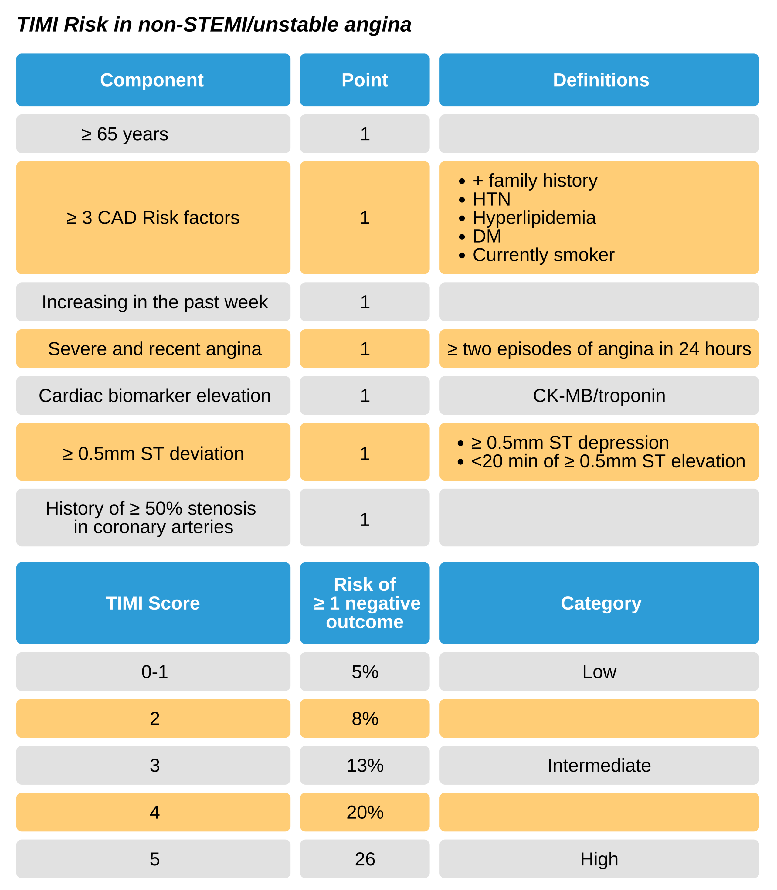
The stratification of non-STEMI patients into these three risk categories is prognostic of outcomes and can guide treatment decisions. This risk score should only be used in patients who have at least an intermediate chance of CAD as the underlying mechanism of chest pain or angina equivalents. Consequently, it may be able to guide the most appropriate treatment for non-STEMI patients. In addition to the TIMI risk score, there is also the TIMI grading system, which evaluates the amount of coronary blood flow from none (0) to complete (3) following revascularization and can be used as an outcome measurement of intervention for ACS.
ST depression and Inverted T waves
These two ECG findings confer high-risk status and indicate early invasive treatment is appropriate.
Combining ECG and cardiac biomarker information
Cardiac biomarkers should be used to help guide treatment alongside ECG changes
- Initial elevation of troponins suggests non-STEMI and high-risk status
- Elevation of troponins on subsequent evaluation suggests non-STEMI and high-risk status
- Negative troponins and ST depression suggests unstable angina
- Q waves can develop in patients with elevated troponins
- Normal or nonspecific ECG changes and elevation of troponins suggest non-STEM and high-risk status.
Management Strategies: Additional therapies
ACS patients with high risk will receive antiplatelet and antithrombosis treatments. Research is continuously updated, and consequently, responders must keep up to date with information and make sure to tailor treatment to the individual as well as follow protocol and consult cardiology as needed.
Alternative treatment includes aspirin, adenosine diphosphate antagonists (i.e., clopidogrel), heparins, GP IIb/IIIa inhibition, IV nitroglycerin for persistent chest pain alpha-adrenergic receptor blockers.
Aspirin
160-325 mg daily aspirin is usually given to all ACS patients unless there is a major contraindication. However, there are no data showing a benefit in non-STEMI/unstable angina subgroup. If patients are following the early intervention treatment, provide the 81 mg dose instead. Continue this dose for the placement of a coronary artery stent.
Clopidogrel
This medication has been shown to have a specific benefit in the non-STEMI/unstable angina group. In those with ACS symptoms and either ischemic ECG changes or elevated cardiac biomarkers, clopidogrel (also heparin and aspirin) within 4 hours of presentation reduced stroke as well as other major adverse events. Additionally, when given at least 6 hours before PCI in non-STEMI patients, adverse events were reduced over the next four weeks.
There is no consensus on the best timing to administer clopidogrel. Some research indicates good benefit at over 12 hours before PCI, while other research indicates that there was less benefit when the dose was given over 15 hours before PCI.
There is some research indicating that clopidogrel is not associated with more bleeding than aspirin alone.
Prasugrel
This medication has been associated with a reduction in non-fatal adverse events, without the mortality benefit of clopidogrel. Additionally, there is an increased risk of major bleeds in non-STEMI patients managed with PCI. Patients with a stroke or TIA history, older than 75 years, or under 60 kg were all at increased risk. Dosing is 60 mg and then 10 mg for maintenance.
Typically, prasugrel is reserved for use as a clopidogrel alternative in
- High and intermediate-risk patients undergoing invasive treatment
- PCI patients.
When the decision is made to perform PCI after angiography, patients should receive aspirin, initial adenosine diphosphate antagonist (if not already given), and stop anticoagulation following intervention unless there is a complication.
Ticagrelor
This is another alternative to clopidogrel for ACS patients undergoing early invasive treatment.
GP IIb/IIIa inhibitors
These medications also block platelet aggregation but at the final common step (different in action from adenosine diphosphate antagonists and aspirin). GP IIb/IIa inhibition can reduce ischemia following plaque propagation. In non-STEMI patients, these medications show benefit in combination with heparin and aspirin when PCI is used. There is an increased risk of bleeding (outside of the brain); however, it is felt that the benefits outweigh the risks in the high-risk group. The same is not true for patients who do not receive PCI; research is mixed in regards to a benefit in mortality and only a slight benefit for recurrent ischemia. Importantly GP IIb/IIa inhibition requires coadministration with heparin. Abciximab, alone of the GP IIb/IIa inhibitors, is not recommended in patients who will not get PCI as research indicates no benefit to these patients.
Recommendations are as follows:
- GP IIb/IIa inhibition is an option instead of clopidogrel/prasugrel for high-risk non-STEMI patients undergoing PCI. Eptifibatide/tirofiban is preferred over abciximab.
- High-risk non-STEMI patients already managed with heparin and clopidogrel can receive GP IIb/IIa inhibition at PCI
- Administer GP IIb/IIa inhibition to patients on clopidogrel/prasugrel and aspirin who have recurrent instability, ischemia or arrhythmia
- Use GP IIb/IIa inhibition in conjunction with aspirin and clopidogrel/prasugrel in high-risk non-STEMI patients
- Use GP IIb/IIa inhibition in patients managed conservatively initially.
Do not administer GP IIb/IIa inhibition to low-risk patients (TIMI score under 3) or at high bleeding risk. In non-STEMI patients who are managed conservatively with no plan for angiography:
- Maintain life-long aspirin
- Maintain clopidogrel/prasugrel for up to a year
- Stop GP IIb/IIa inhibition (if provided)
- Maintain unfractionated heparin for 2 days or LMWH for at most 8 days or hospital length, then stop
There are certain contraindications associated with GP IIb/IIa inhibition:
- Do not use if recent bleeding in the last month or bleeding diatheses; ensure platelets above 150K
- Do not use if a positive 30-day history of intracranial bleed, cancer, aneurysm, arteriovenous malformations, stroke, major trauma or surgery
- Do not use if significant hypertension, pericarditis or aortic dissection
- Do not use if allergic
Heparin
- LMWH or unfractionated heparin can be used in non-STEMI patients who will be managed conservatively
- LMWH or unfractionated heparin can be used in non-STEMI patients who will be managed with early intervention
- Fondaparinux and unfractionated heparin (must be co-administered) use in PCI does not appear to improve outcomes compared to unfractionated heparin alone
- Both bivalirudin and unfractionated heparin can be used in non-STEMI patients with renal failure
- Bivalirudin, fondaparinux, and unfractionated heparin can be used in a non-STEMI patient at risk for bleeding when anticoagulation is indicated
- No research indicates the benefit of anticoagulation outside of the hospital in non-STEMI patients
Managing Angina
IV Nitroglycerin
Nitroglycerin causes dilation of the coronary vessels improving blood flow to the obstructed areas while reducing oxygen needs. However, there is no evidence of long-term benefits and no evidence for using IV, topical or oral doses. Importantly nitroglycerin in the setting of hypotension can lead to worsening of hypotension and may preclude the use of other more beneficial medications (i.e., Beta-blockers/ACEI).
IV Nitroglycerin can be used for:
- Pain resistant to three doses of sublingual/spray/topical nitroglycerin
- Recurrent chest pain
- In combination with beta-blockers to manage hypertension
- In CHF associated ACS

Intravenous nitroglycerin manages angina.
Beta-blockade
Beta-blockers inhibit the sympathetic nervous activity and therefore decrease heart rate and contractions while causing vasodilation and reducing afterload. This is very helpful in ACS as it minimizes infarct, limits ischemia, and decreases arrhythmias. Beta-blocker should be given at the time of presentation in the ED unless there are contraindications
Analgesics
There is data that morphine actually worsens outcome in non-STEMI patients. It is not recommended.
Statins
These medications confer benefit (reduction in recurrent infarct, arrhythmias, and angina) when given within days following ACS onset. 80mg of atorvastatin at 12 hours before and 40 mg at the time of PCI in non-STEMI patients decreases 30-day mortality, death, and urgent vascular intervention.
Cardiac Angiography
Angiography allows indirect visualization of the occlusion of coronary arteries. This diagnostic procedure can then become treatment as angioplasty at the time of angiography can dilate any areas of occlusion. Additionally, cardiac stenting can be done at the same time to ensure continued perfusion. This is usually the case for patients with significant obstruction in one (or sometimes two) vessels. Patients with multiple vessels with obstruction will likely require surgery (coronary artery bypass/CABG).
Revascularization
Both PCI and CABG allow revascularization of obstructed arteries and can reduce adverse events and recurrent disease. CABG is used for disease int the left main coronary, multiple vessels with obstruction, or if PCI cannot be done.
Patients with reduced renal function
- Estimate renal function using creatinine clearance and adjust medications as needed
- Provide necessary IV hydration if contrast material will be used. Also, identify the upper limit of contrast material that can be safely used by measuring the volume of contrast to the renal function ratio. This can help decrease subsequent nephropathy
- Invasive treatment can be provided to patients with mild or moderate (stage II and III) renal disease; however, it is not known if the benefits outweigh the risk in patients with higher-grade renal disease.
Other recommendations
- In non-STEMI patients can be genotyped for loss function CYP2C19 variants if this will help guide treatment
- Insulin can be used for blood glucose management in non-STEMI patients. Maintain glucose under 180 mg/dL and ensure no hypoglycemia.