Manual Defibrillation
To effectively stop VF, enough electrical energy is required. Too much energy runs the risk of causing cardiac muscle damage and may lead to cardiac dysfunction. It is useful to understand the basics of defibrillation to avoid this potential complication.
Defibrillation passes electrical current (charged particles or electrons) through the cardiac muscle in a short amount of time. Electrical flow is known as current, and amperes (A) is the unit of measure. Pressure is required to push the current, and this pressure is known as voltage with volts [V] as the unit of measure. There is always a certain amount of resistance to the current, which is measured in ohms (σ). Generally, defibrillation will end VF within 400–500 milliseconds (ms) of shock delivery.
Understanding the Components of Electrical Shock (Defibrillation)
Missing Table
How does defibrillation occur?
Defibrillation occurs because of the flow of energy or current. While responders will choose an energy for defibrillation, more is needed to move the current to the heart muscle. The amplitude of the current and its change over time, as well as the duration of the shock, will determine the quality of the defibrillation.
The significant factor is the current density, or the amount of current over a given area of the heart muscle measured in amperes/cm2. This concept is important in understanding monophasic vs. biphasic defibrillation. Current density depends on the shock dose, while the fractional trans-cardiac current itself is independent of the dose. Successful defibrillation depends on the location of the defibrillator pads and the anatomy of the chest.
Transthoracic Resistance
Ohm’s law represents the interaction among current, voltage, and resistance. Based on this interaction, responders can directly impact resistance more than the other two factors.
Transthoracic resistance depends on energy, pad size, skin-to-pad contact, coupling materials, pad pressure, frequency of shocks, ventilatory phase, and chest size. While there is a range of transthoracic resistance in patients, the average adult has a resistance between 70 and 80σ. A high resistance decreases the current’s ability to affect the heart.
Requirements for Defibrillating VF
Successful defibrillation requires enough current to flow to the heart and stop the aberrant cardiac arrhythmia (VF) but not so much current that it would cause cardiac muscle damage. It has to be just right.
By selecting the correct current, responders can eliminate the need to give multiple shocks as well as limit the risk of cardiac muscle damage. The correct energy dose is really based on the fractional trans-cardiac current, which depends on the location of the defibrillator pads in relation to the heart as well as any resistance to the current between the defibrillator pads. This interaction will determine the overall current density and the efficacy of defibrillation. Surprisingly, chest size plays a rather small role in this interaction.
Types of Defibrillation Waveforms
All defibrillators currently used provide electrical current via waveforms: either monophasic or biphasic. While the specifics may differ based on manufacturer and device, the two basic waveforms have stable characteristics.
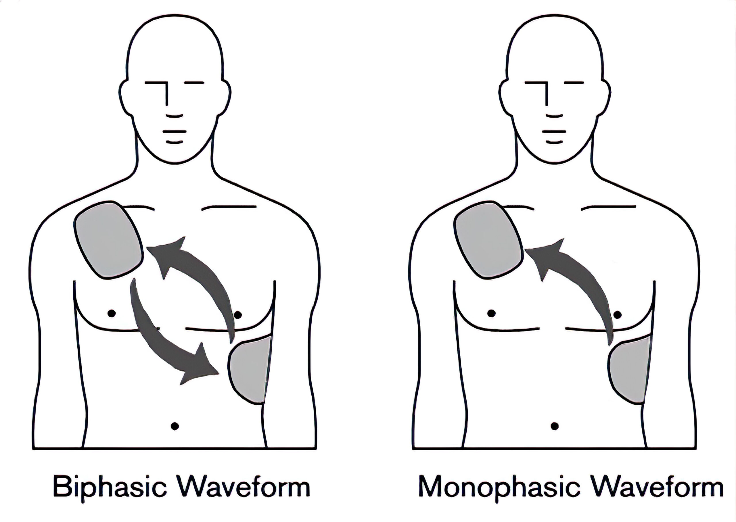
Monophasic waveforms push current in mostly one direction. Biphasic waveforms push current in one direction for some time, then switch direction for the remainder of the time. The first part of the biphasic waveform prepares cardiac cells for effective depolarization.
Additionally, defibrillation can vary the rate of change of the waveforms. For example, a dampened sinusoidal waveform rises quickly. It dissipates slowly while a truncated exponential wave rises quickly then cuts off quickly. Most monophasic defibrillators in use produce dampened sinusoidal waveforms.
Biphasic defibrillators tend to be more effective for implantable devices. While the exact reason for this is controversial, the biphasic waveform can defibrillate the cardiac muscle using less current than the monophasic defibrillator. This is useful for transthoracic defibrillation as the factors affecting current density (such as pad location and chest anatomy) are not well defined.
However, biphasic defibrillation does not guarantee a higher rate of ROSC or survival after cardiac arrest. Consequently, both types of defibrillators can be used for treating atrial and ventricular arrhythmias. (Class I, Evidence level B-NR). However, due to the improved termination of arrhythmias, biphasic defibrillation should be preferred over monophasic defibrillation for atrial and ventricular arrhythmias. (Class IIa, Evidence level B-NR).

Biphasic and Monophasic Defibrillation Waveforms
Generally, for terminating VF, responders should follow the manufacturer’s instructions to choose the dose of energy for the initial shock. If there are no instructions, the maximal energy dose available should be considered. (Class IIb, Evidence level C-LD).
Monophasic Defibrillators
Currently, not many monophasic defibrillators are being produced, although many are still available for use. These defibrillators provide current in one direction or polarity. They are categorized by the speed at which the current drops to zero. Monophasic with damped sinusoidal (MDS) defibrillation slowly drops to zero while monophasic with truncated exponential waveforms drops to zero suddenly.
MDS is the most common monophasic defibrillation waveform in use. When using monophasic defibrillation, the initial shock is 360 J, as are the subsequent shocks if VF is not immediately corrected. Following shocks, chest compressions should be resumed immediately with a heart rhythm check only after 2 minutes of CPR.
The interval between chest compressions and defibrillation should be as short as possible as just a few seconds can improve the chance of successful defibrillation and subsequent ROSC. It is important to improve the coordination among responders providing chest compressions and those delivering defibrillation. Practicing clearing the patient and the quick delivery of a shock is a vital method for achieving this goal. Rescue breathing can be eliminated before providing defibrillation as this can delay time to defibrillation.
Biphasic Defibrillators
Research evaluating defibrillation success both inside and outside of hospitals indicates that low energy biphasic defibrillation has a similar or improved rate compared to monophasic defibrillation using increasing energy doses (beginning at 200 J and increasing to 300 J, then 360 J). There has been no direct comparison of the different types of biphasic defibrillation.
The optimal energy dose for biphasic defibrillation is not known. But research indicates that a biphasic energy dose ≤ 200 J has a similar or improved rate of success at correcting VF when compared to monophasic defibrillation or higher energy doses. Responders should follow the instructions from the manufacturer to choose the best dose of energy for defibrillation.
Patient differences in terms of resistance can be managed by manipulating the length and voltage of defibrillation as well as by releasing any remaining membrane charge (burping). The best parameters for the duration of the waveform phases are not known. Additionally, there is no evidence that one type of biphasic waveform improves either immediate-, short-, or long-term outcomes. It is most likely that other patient factors, such as time to defibrillation and chest compressions, have a more significant role in affecting outcomes.
The recommended dose for biphasic defibrillation is generally between 120 J and 200 J. If there are no instructions by the manufacturer, the maximum available energy dose should be chosen. If biphasic defibrillation is unavailable, monophasic defibrillation may be used.
Generally, energy doses remain constant or escalate with subsequent shocks. Again, while there is no evidence regarding the best dose for biphasic defibrillation, there is no evidence in human studies for detriment with doses as high as 360 J. However, in animals, higher doses than 360 J have been shown potentially to cause cardiac damage. AHA recommendations indicate that subsequent defibrillation energy doses should at least be maintained and can be increased if this is an option.
There is some new evidence that using current, rather than energy, dosing may be more effective in monophasic defibrillation. There is no evidence yet indicating that this is the case in biphasic defibrillation, but this innovation is under investigation and may become the new standard of care.
Placing the Defibrillation Pads
A very important but often forgotten consideration is the placement of defibrillation pads and paddles. Their location affects resistance, fractional transcardiac current, and current density in the cardiac muscle. Accurate placement of paddles leads to only 4–25% of the delivered current reaching the cardiac muscle.
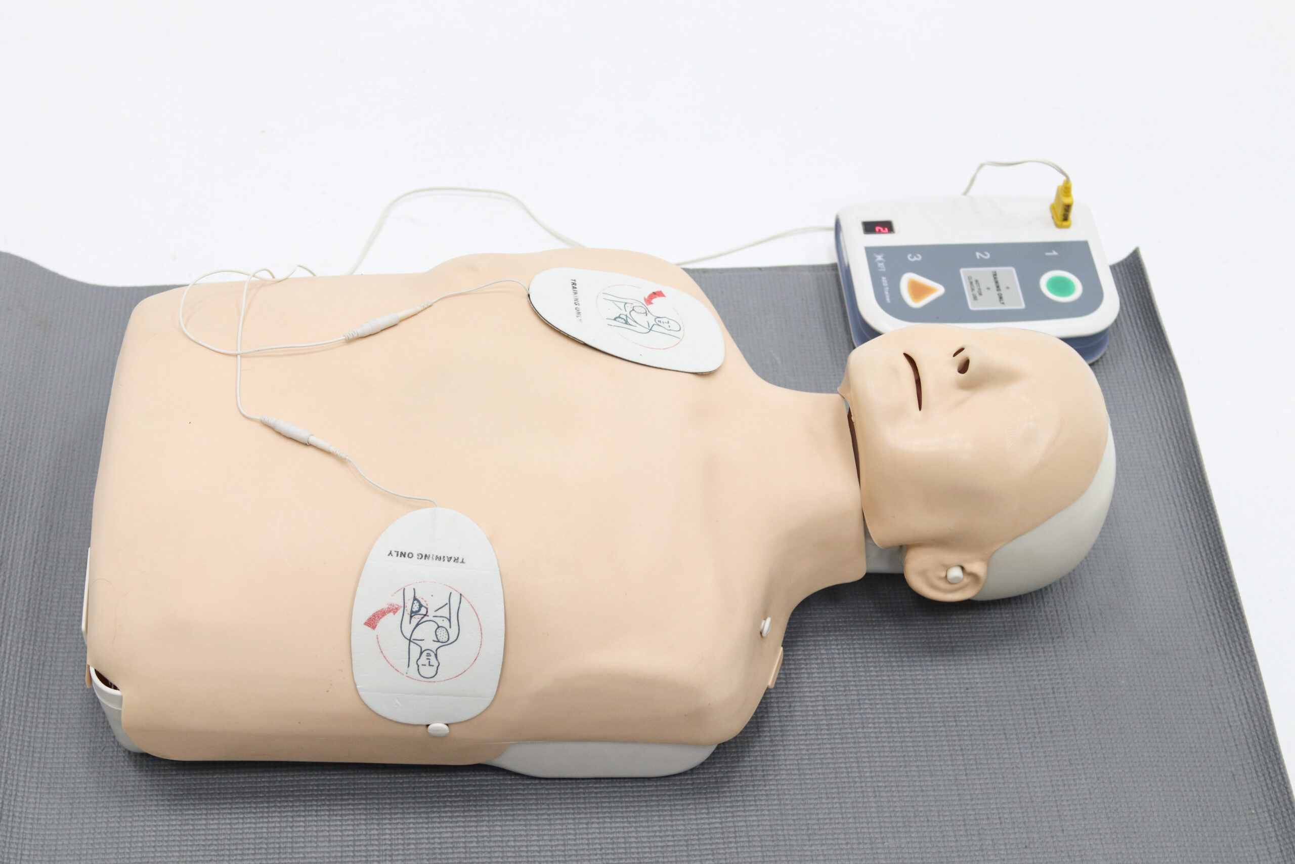
Pads and paddles are best placed in either the sternal-apical or the anterior apex configuration. The first pad should be placed on the anterior chest at the right side of the sternum, just inferior to the clavicle. The other pad should be placed in the middle of the left axillary line on the left side of the nipple. An alternative option is to place the first pad over the apex of the heart (left precordium) and the other pad below the scapula on the left side of the back. Both options improve the current flow through the cardiac muscle and increase the chance of defibrillation success. The anterior placement is often more convenient, but both are acceptable depending on preference and patient circumstances.
It is common to place the defibrillation pads very close to one another. When pads are placed too close, the current can miss the cardiac muscle. Additionally, if the anterior pad is placed over the sternum, rather than to the right of it, the sternum will block a significant amount of the current.
The pads or paddles used for adults should be between 8 and 12 cm. However, pads on the larger end of the spectrum may be more effective. Smaller pads can damage the heart by causing cardiac muscle necrosis. Responders should always ensure the pads are fully in contact with the skin. Additionally, when treating arrest in pediatric patients, the correct size must be used as an overly small size can lead to increased transthoracic resistance.

Anterior Placement of Defibrillator Pads
Defibrillation in Patients with ICDs or Pacemakers
Occasionally responders will need to defibrillate an individual with an ICD or implanted pacemaker. Responders must not place the pads over or close to the pacemaker or generator, as this can cause malfunction of the device. Additionally, the ICD or implanted pacemaker can block current flow to the cardiac muscle, decreasing the likelihood of successful defibrillation.
Key Takeaway
Do not place defibrillation pads over any hard lump in the chest or any medication patch.
In the past, the general recommendation was to keep pads at least 2.5 cm or 1 inch away from the implanted device. A contemporary study evaluating external cardioversion for atrial fibrillation in patients with ICDs or pacemakers showed that placement of the pads at least 8 cm or 3 inches from the device was safe and did not result in damage to the implanted device. All implanted devices were in the infraclavicular region, and the pads were placed in the anterior-posterior position. Additionally, the current flow was perpendicularly oriented to that of the device’s implanted leads. Both biphasic and monophasic delivery were performed with shock intervals of at least 5 minutes.

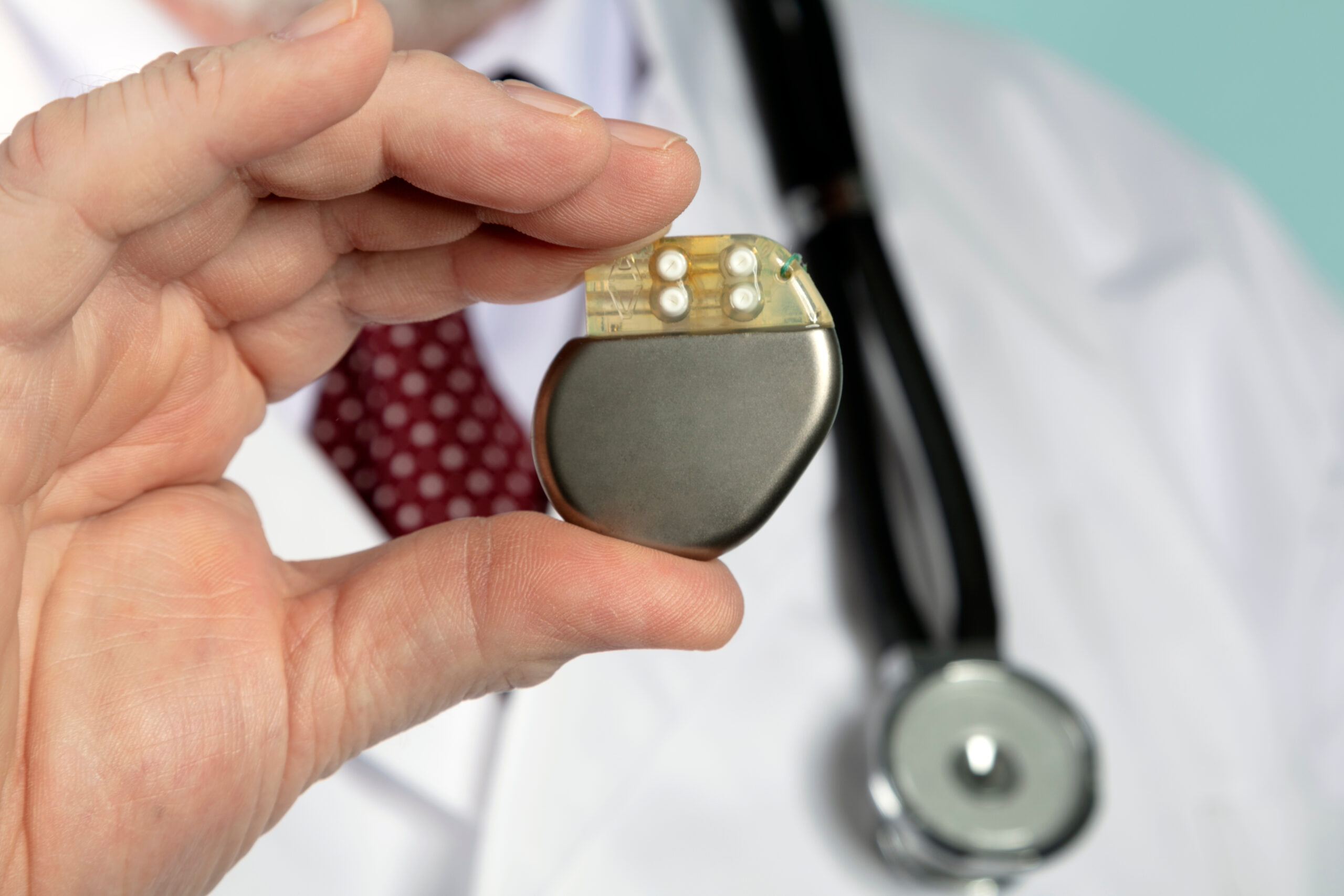
A pacemaker is usually implanted under the skin in the chest.
While the management of the ICD is generally the same as implanted pacemakers, it is important to note that there are patients with ICDs placed in the abdomen rather than in the chest. In this case, the traditional placement of the pads is satisfactory if there is enough distance from the generator. Higher energy doses may be needed in these situations due to increased cardiac insulation from the epicardial defibrillation electrodes.
If the implanted ICD provides shocks to the patient (visualized by muscular contractions), responders should wait between 30 and 60 seconds to attach an external defibrillator. Generally, ICDs can complete therapies for arrhythmias within the first few minutes after collapse. Consequently, if an unconscious patient with an ICD is found, the ICD has likely completed all available therapies.
This patient should be treated as a cardiac arrest patient without an ICD, except for noting its location and ensuring a safe distance of 8 cm between the ICD and the defibrillator pads. Although ICDs tend to be less affected by external defibrillation than implanted pacemakers, they can still be damaged by defibrillation.
Analysis of the cardiac rhythm and shock delivery of the ICD and AED can conflict. Consequently, rapid-fire shock can occur as both detect an arrhythmia that can be shocked. In this case, responders should be quick to stop the external defibrillation after recognizing patient contractions, indicating ICD defibrillation. Since the ICD can pace bradycardia and tachycardia, this can erroneously lead the external defibrillator to be unable to detect an arrhythmia that can be shocked. In this case, the manual defibrillator mode can be used to allow responders skilled in cardiac analysis to detect shockable rhythms.
Using the AED in the Hospital
AEDs in the hospital setting may improve the patient’s chance of surviving to discharge when treating VF or pVT. Still, in patients with sudden cardiac arrest who are not on telemetry, defibrillation even in the hospital can be delayed for several minutes or longer. A study looking at almost 12,000 patients with cardiac arrest in US hospitals showed that AED use did not improve discharge survival in cardiac arrest patients. There was actually a lower survival rate in cardiac arrest caused by unshockable rhythms in the study.
Additionally, in this study, there was no benefit seen with AED use in shockable rhythms. It was thought that this larger study was more accurate as others had relatively small sample sizes. Regardless, in the hospital, the goal time from cardiac arrest to shock should be 3 minutes or less.
Synchronized Cardioversion
This form of energy shock synchronizes delivery with the QRS wave. Therefore, shock delivery does not occur during the relative refractory period, which can increase the risk of producing VF. Synchronized cardioversion is used for supraventricular tachycardias such as atrial fibrillation and flutter, atrial tachycardia, and reentry SVT.
Pacing
Pacing is used in patients who are unresponsive to atropine and other second-line treatments (if attempting to use them first does not delay care). Pacing should not be used for cardiac arrest due to asystole, as this can delay effective chest compressions. Pacing can be transcutaneous or transvenous.
Key Takeaway
Do not attempt to pace a patient in cardiac arrest due to asystole.
Defibrillator Safety
Safety is key while using defibrillation devices. The pads should never be touching. Overlapping pads or using excessive gel can allow the current to flow onto the chest wall rather than reaching the cardiac muscle.
Generally, adhesive pads are recommended over paddles as pads can be placed before the arrest and do not require additional setup at the time of use. Pads should never be placed on top of transdermal patches that infuse medicines. These patches can block current flow and lead to burns on the skin. If possible, these patches should be removed and any medcation wiped off.
It is important to remember that a cardiac arrest patient lying in water should be moved or the chest dried if it is wet. A patient in ice or snow does not need to be moved. Any excessive chest hair should be quickly removed by applying and quickly removing a pad or by shaving the area. These actions should not delay compressions or defibrillation.
Safely using the Defibrillator in Patients with ICDs
The ICD is unable to provide as much of a shock energy dose as a defibrillator. ICD shocks range between 30 J and 40 J. A fraction, up to one-fifth of this energy, does travel to the patient’s skin and can be felt by others in contact with the patient at the time of shock. Generally, this is too low an energy to cause harm. Responders in contact with the patient in at least two areas and when a conductor (e.g., gel) is used are most likely to feel the shock from the ICD defibrillation. This is usually not the case for the responder providing chest compressions, especially if wearing gloves, which would further insulate them from shock.
Fire Safety with Defibrillation
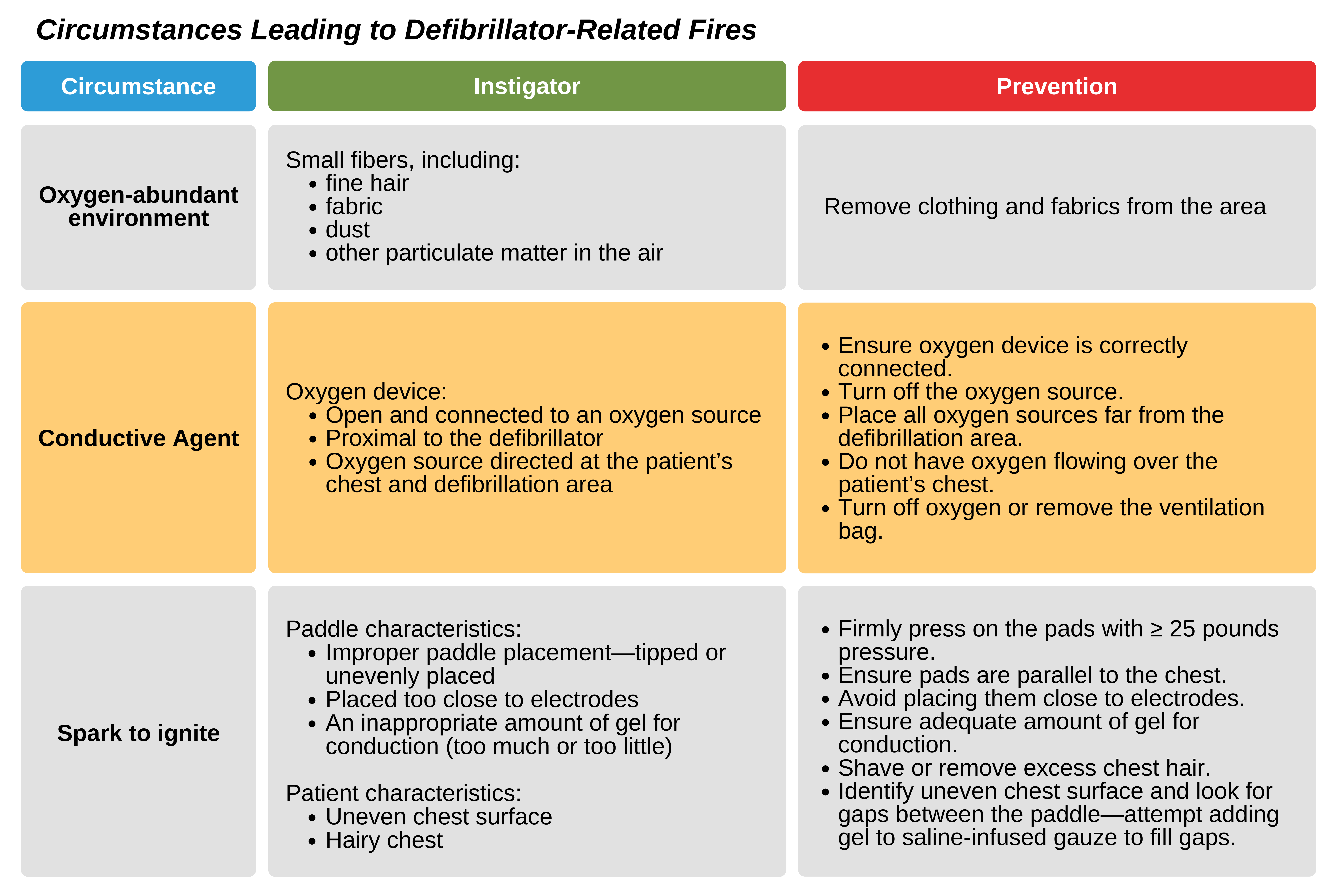
There can be a concern for a fire occurring due to sparks from improperly applied paddles when oxygen is abundant. This is more likely in cases where there is oxygen blowing across the patient’s chest, such as in the case of open ventilator tubing connected to oxygen. Therefore, the responder should try to minimize excessive oxygen flow over the chest during defibrillation.
Adhesive defibrillator pads decrease the likelihood of sparks forming. Generally, when paddles are used, gel pads are recommended over pastes or gel as these increase the risk of sparks. Ultrasound gel or other medical gels that have low conductivity should never be used.
Fires from defibrillation are rare and completely preventable as they require an abundant oxygen environment, a conductive agent, and a spark to ignite. Fires can be prevented easily by preventing an error in defibrillation or oxygen supply.

There is some concern about blanket recommendations to disconnect or turn off the oxygen source before defibrillation. This is because of the theoretical delay in defibrillation caused by taking this step to prevent a fire. If responders use proper defibrillation safety steps, there will be no need to take extra steps to prevent fires. However, patients who are at increased risk should always have oxygen sources removed.
Key Takeaway
Defibrillation should be done in conjunction with high-quality CPR. This means reducing any delays in chest compressions both before and after delivering shocks.
Maintaining Defibrillation Devices for Prompt Use
Defibrillation devices should always be ready for use. This includes ensuring adequate power supply. Generally, most AEDs do routine self-checks to ensure the device readiness. Staff should use a checklist to ensure that everything needed for defibrillation is functioning and maintain the devices according to manufacturer’s guidelines.