Chapter Progress
0% Complete
Get Neonatal Resuscitation Certified Today
Anatomic Landmarks
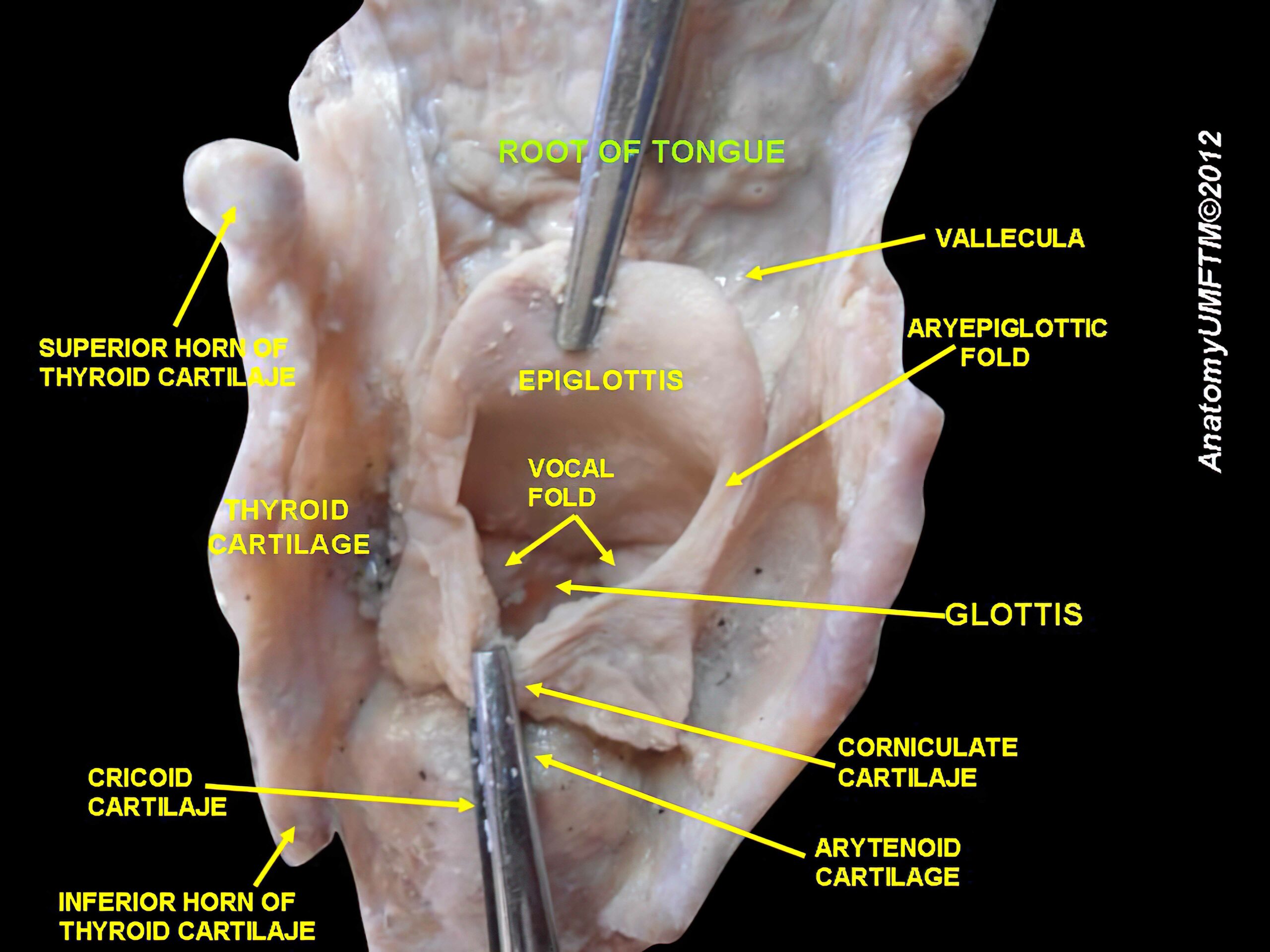
The team member(s) responsible for placing an advanced airway in a neonate must be familiar with these anatomical landmarks in the airway:
- Vallecula: a pouch located at the base of the tongue and the epiglottis. The laryngoscope blade can be inserted into the vallecula and lifted to expose the glottis and vocal folds.
- Epiglottis: a flap of elastic cartilage that covers the glottis. One technique for endotracheal intubation involves guiding the laryngoscope blade toward the epiglottis, then lifting the epiglottis to expose the glottis and the vocal folds.
- Larynx: an anatomical segment of the upper airway that connects the pharynx and the trachea.
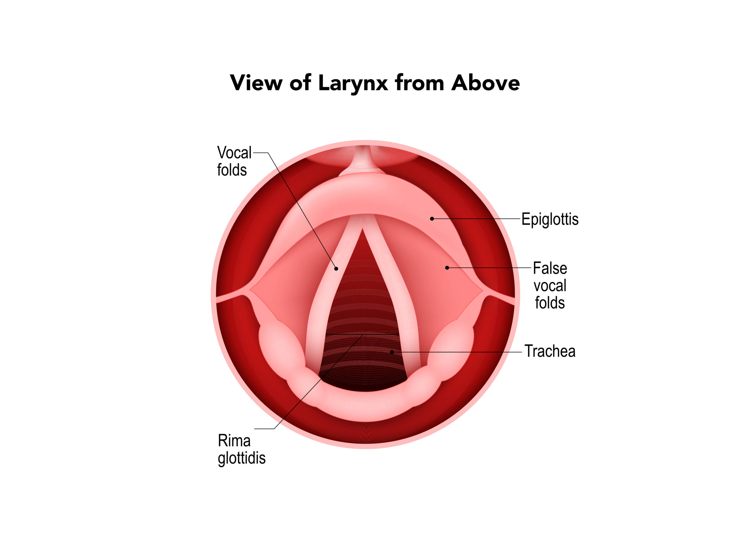
- Glottis: an opening in the larynx that amounts to a passageway into the trachea. The vocal cords line each side of the glottis.
- Vocal cords or vocal folds: two membranous tissues that project medially from the walls of the larynx and form a slit across the glottis. The edges of the vocal folds vibrate when an airstream passes through, causing modulation to produce the voice.
- Thyroid cartilage: a thick cartilage that surrounds and protects the larynx.
- Trachea: the segment of the airway located between the larynx and the carina.
- Carina: the junction between the two main bronchi.
- Main bronchi: the bifurcated passageway that delivers air to the right and left lungs.
- Esophagus: a passageway that is anterior to the trachea and leads to the stomach.

Introduction to The Larynx (Posterior View)

View of the larynx from above.