Umbilical Venous Catheterization
Chapter Progress
0% Complete
Get NRP Certified Today
Umbilical Venous Catheterization
Prior to each delivery, the team ensures that all the necessary equipment is available for umbilical catheterization. Prepackaged kits contain all the necessary components for a successful procedure.
If a prepackaged kit is not available, the team should ensure that the following items are available:
- Mosquito hemostat, straight and curved
- Vessel dilator probe
- Iris forceps, half-curve
- Iris forceps full curve
- Iris forceps straight
- Straight iris scissors
- Baumgartner hemostat needle holder
- Straight suture scissor
- 4-0 suture
- Umbilical tape
- 3F or 5F single lumen umbilical catheter
- A 3-way stopcock
- Antiseptic solution
Umbilical venous catheterization requires an aseptic technique.
- The umbilical catheter is filled with normal saline from a 10 mL syringe connected to a 3-way stopcock.
- The stopcock is placed in the “off” position to prevent air from entering the catheter system or loss of fluid.
- The umbilical cord is cleaned with an antiseptic solution.
- Umbilical tape is loosely tied at the base of the umbilical cord.
- Chest compressions are briefly halted, and everyone warned that a scalpel is in use.
- The umbilical cord is cut just below the umbilical clamp, about 2 cm above the skin line. Care is taken to cut across rather than at a slanted angle. This technique exposes the umbilical vein near the 12 o’clock position.
- The umbilical catheter is inserted 2–4 cm into the umbilical vein.
- Free-flowing blood should be visible within the catheter after switching the stopcock to the “on” position.
- The catheter must not be inserted too deeply to prevent the epinephrine from being directly administered to the liver.
- The catheter is held in place with one hand during the resuscitation. A transparent adhesive tape such as Tegaderm® can be used to assist in holding the catheter in place.
- The catheter is removed by gently pulling it. The umbilical tape may be tied or pressure applied to the cord stump to prevent bleeding.
- If the clinician decides to keep the umbilical vein catheter in place after resuscitation, it can be sutured or secured with “goal post” tape.

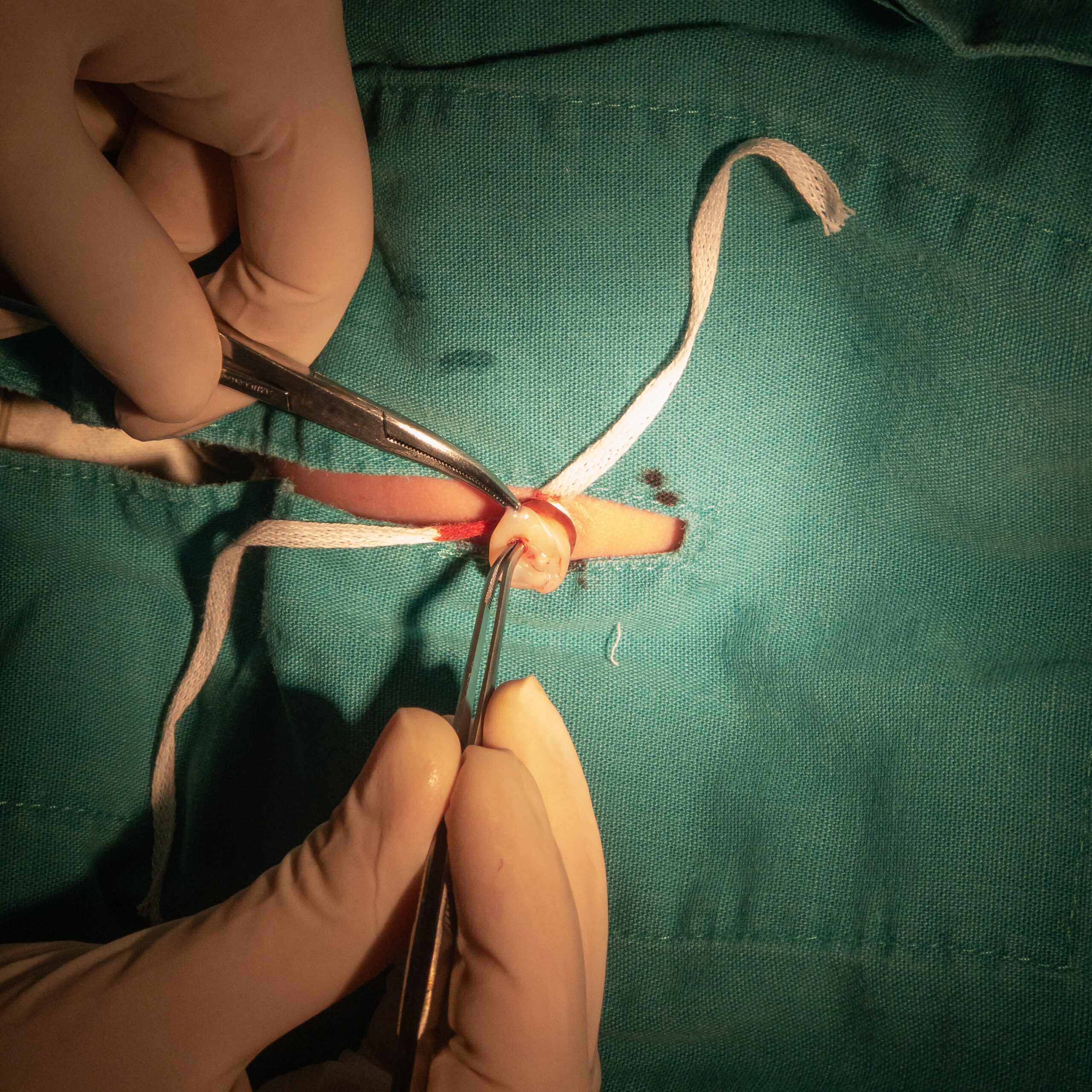
Umbilical cord ready for intravenous catheter placement.

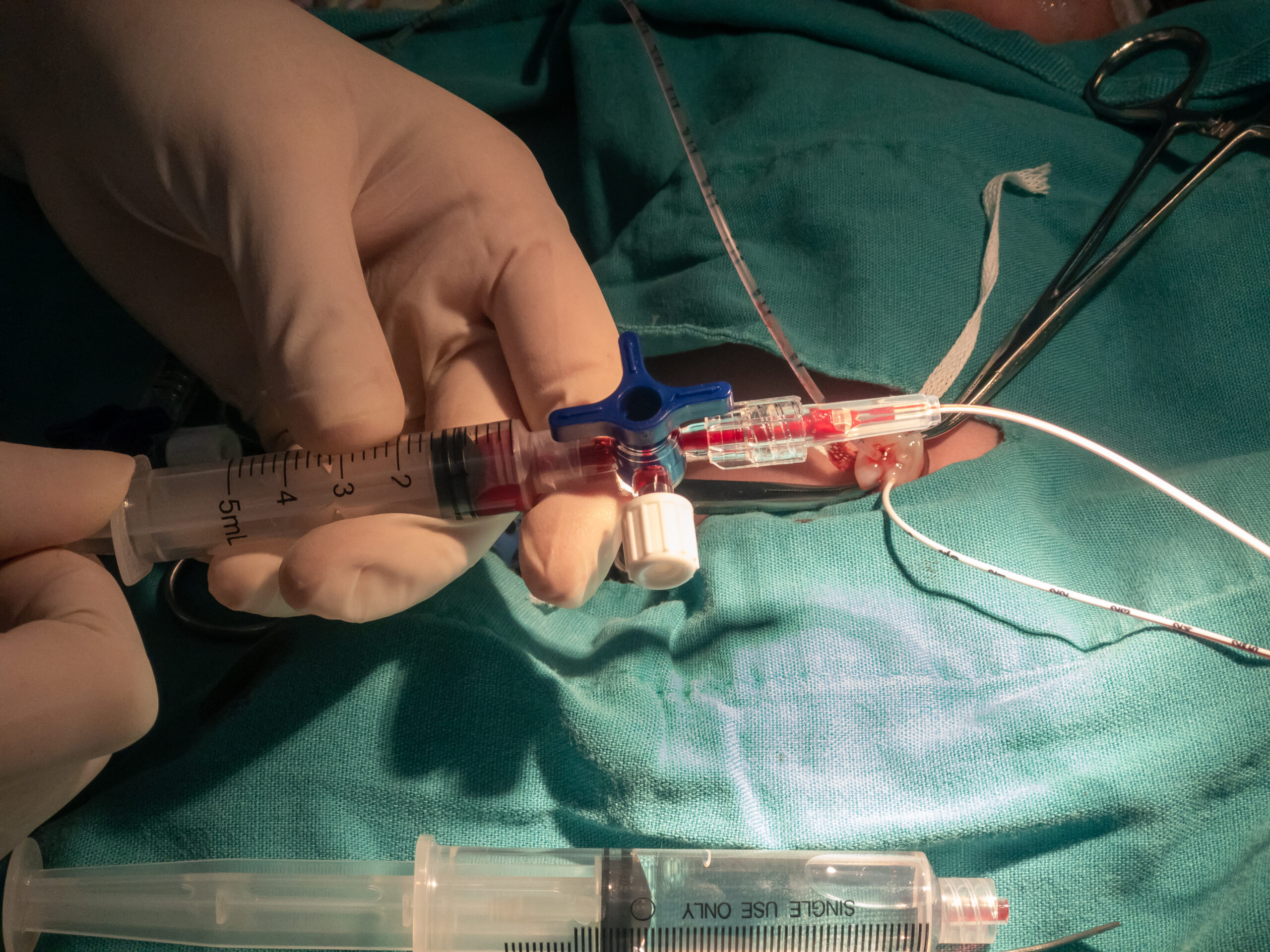
Umbilical venous catheterization.