Anatomy & Physiology of the Fetal Circulation and Transitioning Circulation
Knowledge of fetal circulation and the neonate’s transition from intrauterine to extrauterine circulation at birth guides the healthcare provider on neonatal resuscitation’s basic concepts. In the uterus, the fetal lungs are filled with amniotic fluid and do not participate in the gas exchange of carbon dioxide and oxygen. The mother supplies oxygen via the placental circulation. This fetal circulation path is also known as a right-to-left shunt because blood flows from the right heart to the left heart, bypassing the lungs.
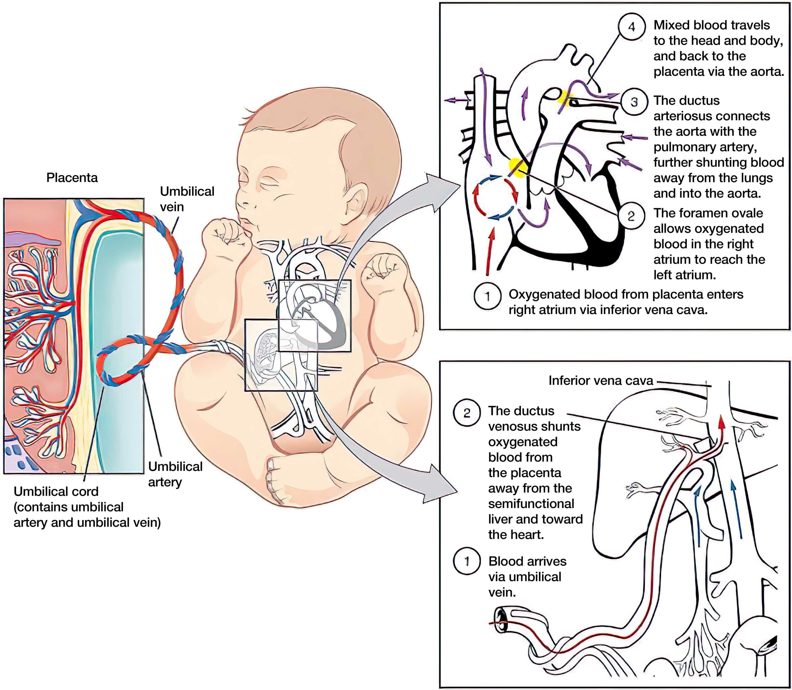
Oxygen from the mother’s blood diffuses into fetal blood vessels at the placenta. Oxygen then travels from the placenta to the fetus through the umbilical vein. The umbilical vein crosses the liver, joins the inferior vena cava, and enters the right side of the fetal heart. Most of the blood bypasses the lungs because the pulmonary arteries and veins constrict.
Blood flows through an opening in the atrial wall known as the foramen ovale and directly to the aorta through an opening in the pulmonary artery known as the ductus arteriosus. Well-oxygenated blood circulates to the rest of the fetal tissues and organs, with the brain and heart receiving the most supply. Deoxygenated blood then returns to the placenta via two umbilical arteries. The carbon dioxide produced from fetal metabolism is transported across the placenta and removed by the mother’s lungs.

Fetal circulation.
Transitional circulation involves a series of physiologic changes and adaptations. At birth, the lungs of the newly born must become the primary organ for gas exchange. The transition from fetal circulation to neonatal circulation begins when the baby takes the first deep breaths and cries outside of the uterus. As the lungs fill with air, the alveoli inflate, and the alveolar fluid dissipates. The presence of oxygen in the alveoli also stimulates the pulmonary blood vessels to dilate, diverting blood flow into the alveoli. Oxygen from the inspired air is absorbed in the pulmonary veins.
At the same time, the metabolic processes of cells remove carbon dioxide from the pulmonary arteries. The foramen ovale and ductus arteriosus close, diverting blood from the right atrium and right ventricle into the pulmonary artery. The fetal right-to-left shunt is no longer present. Oxygenated blood from the pulmonary veins enters the left side of the heart and is then pumped into the aorta and out to the rest of the body.

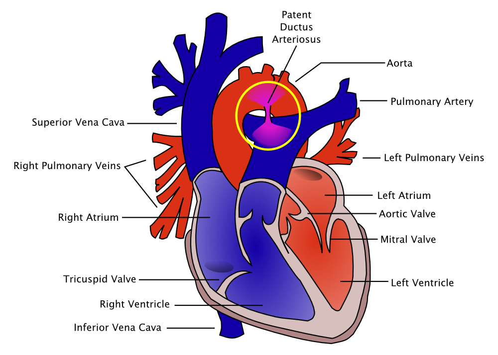
The ductus arteriosus is a fetal artery connecting the aorta and the pulmonary artery. This allows blood to detour away from the lungs before birth.3
The transition process occurs within the first few minutes of birth and completes within several days. The newly born’s oxygen saturation does not reach 90% until the 10th minute of life. It takes time for the alveoli to absorb the alveolar fluid. The complete closure of the ductus arteriosus usually occurs 24 to 48 hours after birth, and the process of fully relaxing the pulmonary arteries may continue for several months.
Disruption in Fetal or Transitional Circulation
When the placental function or transitional circulation is compromised, the decrease in oxygenated blood to various organs triggers a survival reflex. Vessels constrict to divert blood to vital organs such as the heart and brain. The survival reflex is a short-term fix, and if the underlying problem is not quickly corrected, the heart will lose function, and circulation will be compromised. The deprivation of oxygenated blood to the cells and tissues leads to organ damage.
Key Takeaway
When transitional circulation is compromised, a survival reflex diverts blood to the heart and brain.
Clinical indications of an abnormal transition include:
- Irregular respiration, apnea or tachypnea
- Bradycardia or tachycardia
- Decreased muscle tone
- Low oxygen saturation
- Low blood pressure