Targeted Management of Respiratory Distress or Failure
After stabilizing oxygenation and ventilation, the PALS provider identifies the respiratory problem: (1) upper airway obstruction, (2) lower airway obstruction, (3) lung tissue disease, or (4) disordered control of breathing.
Managing Upper Airway Obstruction
The upper airway includes the nose, pharynx, or larynx and other respiratory airways outside the thorax. Common causes of upper airway obstruction include foreign-body obstruction, edema, an acute infection, thick secretions, enlarged tonsils or adenoids, a tumor, or a congenital malformation of upper airway structures.
Poor CNS control can also contribute to upper airway obstruction. In a seriously ill patient with a depressed level of consciousness, the muscles in the pharynx relax. As a result, the tongue may fall back into the oropharynx and cause an obstruction. A newly born infant may have a prominent occiput due to childbirth, and when the newborn is placed supine, the resultant neck flexion may cause obstruction.
General management emphasizes measures that relieve the obstruction:
- Comfortable positioning
- Manual airway maneuvers such as jaw-thrust or head tilt-chin lift
- Removal of any foreign-body obstructions
- Suctioning to remove secretions
- Calming the child to minimize further obstruction caused by crying
Epinephrine and corticosteroids given IV or IM can help relieve swelling in the pharynx. Some corticosteroids can also be inhaled. Noninvasive positive ventilation may help if the child tolerates it without excessive crying.
An oropharyngeal airway can help maintain a patent airway but must only be used with patients who are unconscious with absent gag or cough reflexes. If the patient is conscious, the provider can use a nasopharyngeal airway instead. However, a nasopharyngeal airway can potentially injure a child even further if there is trauma to the face.
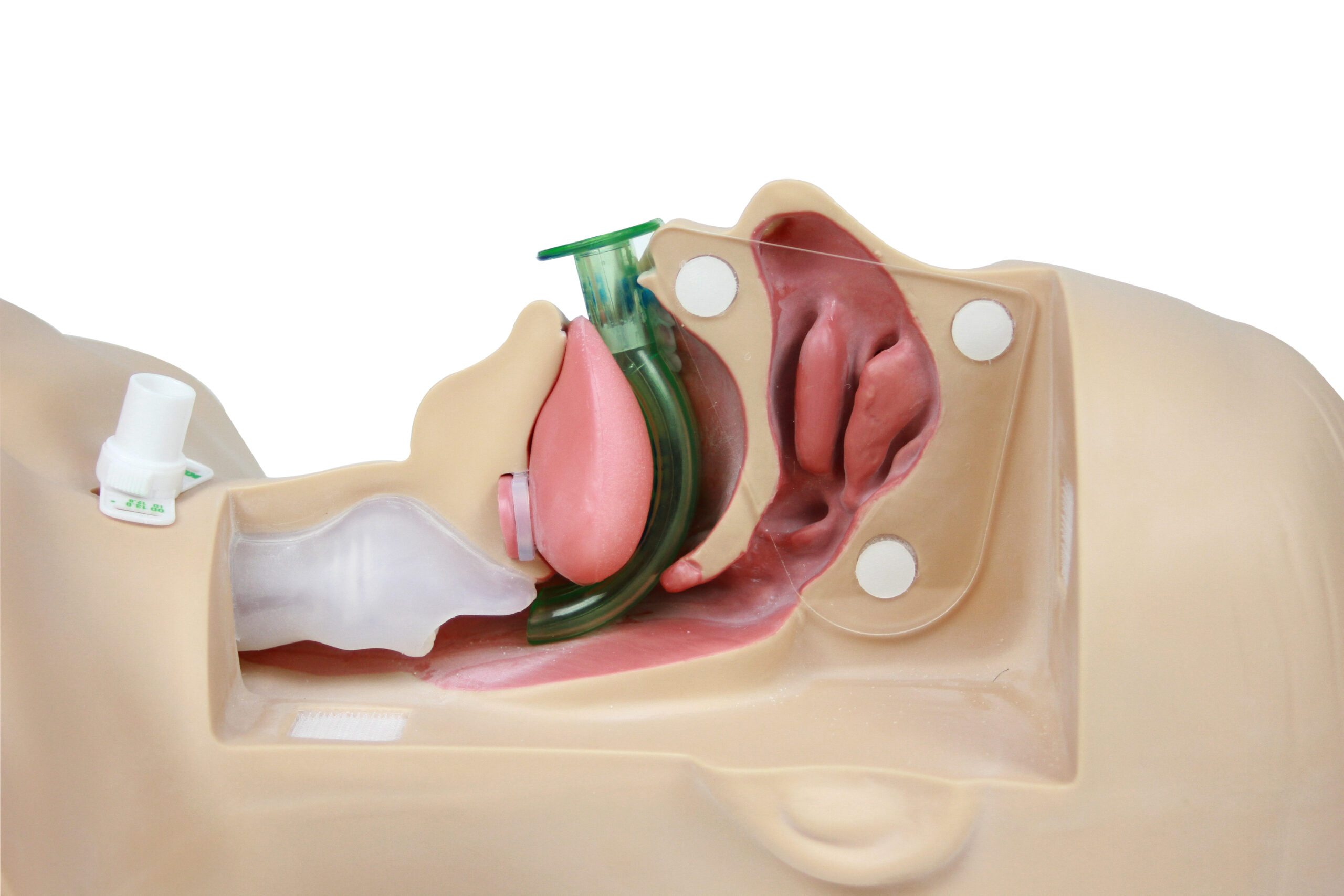
Oropharyngeal Airway Device Placement
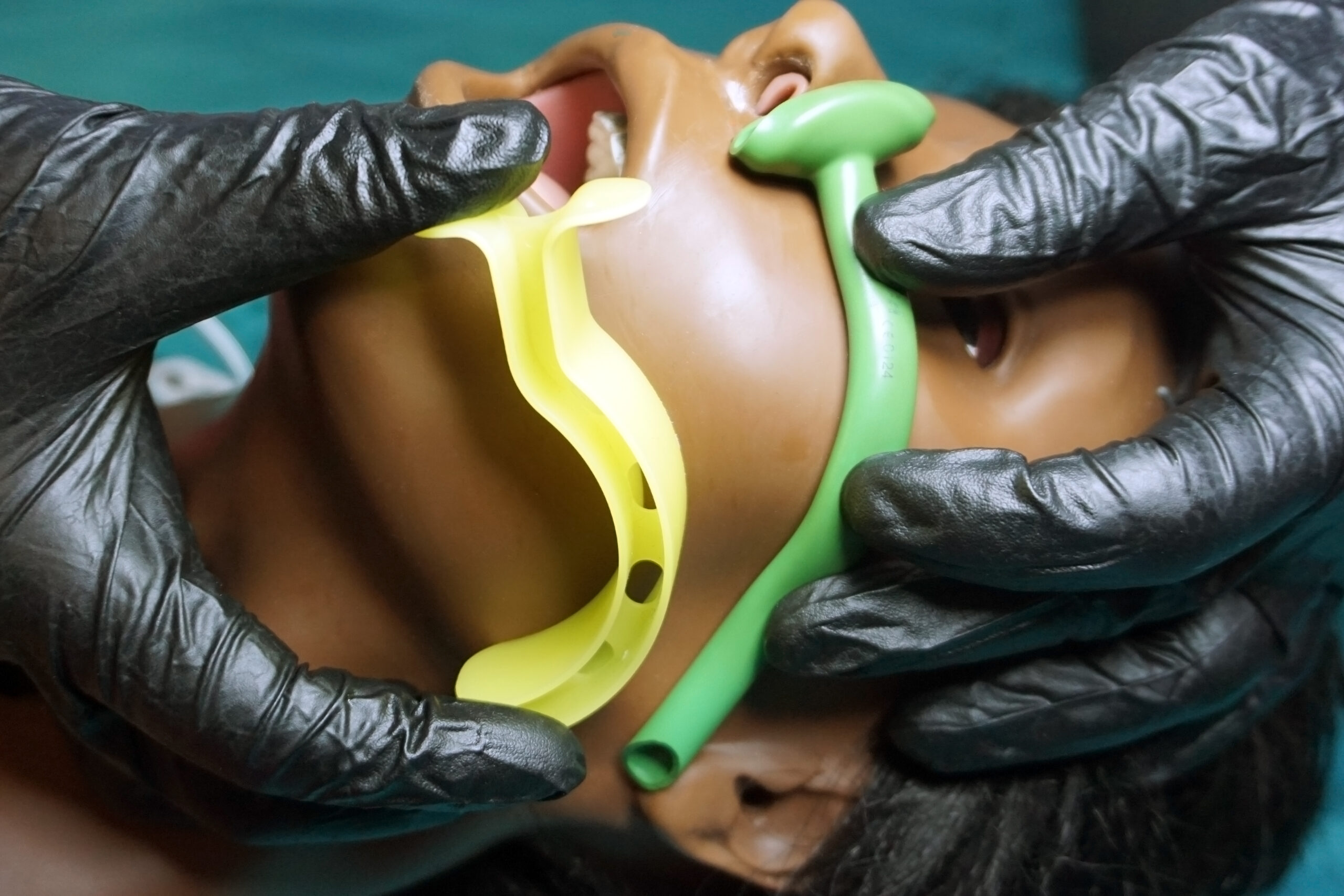
Oropharyngeal Airway and Nasopharyngeal Airway Size Confirmation Method
Croup
Croup is usually caused by a viral upper airway infection. The infection causes edema of the larynx and trachea, which increases airway resistance. Stridor and a seal-like barking cough are common presenting signs of croup. Stridor often worsens when the child cries.
Mild croup is characterized by an occasional barking cough without evidence of stridor or increased respiratory effort and is treated with prednisolone or dexamethasone.
Children with moderate to severe croup have a frequent barking cough, audible stridor at rest, retractions, agitation, and decreased air entry on auscultation. Treatment is with humidified oxygen, nebulized epinephrine, and dexamethasone. If the patient’s status does not improve, heliox (a helium-oxygen mixture) may be considered. The inert gas characteristics of heliox may aid in reducing turbulent flow patterns associated with upper airway obstruction.
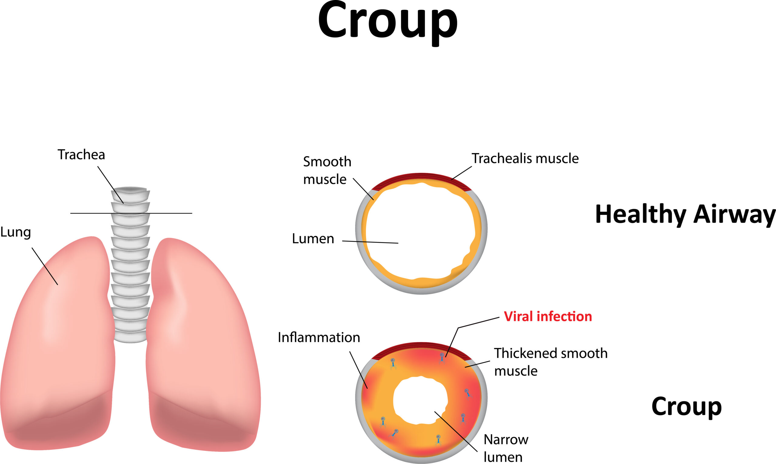
Croup is an upper airway infection that causes a distinctive barking cough.
If the patient with croup deteriorates to impending respiratory failure, the team must administer high concentrations of oxygen via a nonrebreather mask. If hypoxemia is present or changes in sensorium occur despite oxygen supplementation, the team should provide assisted ventilation via the bag-mask device. Dexamethasone should be delivered via IV or IM.
If endotracheal intubation is necessary, an ET tube a half-size smaller is recommended since there is likely swelling of the subglottic area. If the provider is unable to insert the ET tube, an emergency tracheotomy may be needed.
Anaphylaxis
The first step in treating an allergic reaction is to identify and remove the offending agent to prevent it from doing further harm to the child.
If the allergic reaction is mild and there is no upper airway obstruction, treatment of the child with an oral antihistamine such as diphenhydramine may be appropriate. H2 blockers such as famotidine can be considered as an adjunct treatment. However, if upper airway obstruction is apparent, the provider administers an antihistamine IV or IM.
If there is a moderate to a severe anaphylactic reaction, the provider must give epinephrine IM via an autoinjector or a regular syringe once every 15 minutes as needed. An IV corticosteroid such as methylprednisolone should also be considered. If bronchospasm is apparent, albuterol via metered-dose inhaler or nebulizer is indicated. Albuterol delivery can be repeated every 15 minutes as needed or delivered continuously. If respiratory distress is apparent, endotracheal intubation may be needed.
Anaphylaxis can also cause extravasation of fluid that can lead to significant hypotension. In this case, the provider can administer an isotonic crystalloid solution such as normal saline or lactated Ringer. A bolus dose of 20 mL/kg IV should be administered and repeated as needed to increase blood pressure back to normal.
If these interventions fail to treat the patient’s symptoms, an IV infusion of epinephrine may be required. The epinephrine infusion should be titrated to achieve adequate blood pressure.
Foreign-Body Airway Obstruction
The provider should suspect mild foreign-body airway obstruction (FBAO) if the pediatric patient is still able to make sounds and cough forcefully. The child should be encouraged to remove the obstruction by making use of the coughing reflex. Additional emergent intervention is not warranted at this point. The provider should call for help from a second person in case the obstruction worsens.
If the FBAO becomes severe, the child will be unable to make a sound, speak, or cough. There will also be an absence of air exchange. A high-pitched noise might be heard during inhalation or no noise at all.
If the child in severe distress with suspected FBAO nods or otherwise indicates “yes” when asked if they are choking, the rescuer stands behind the child and delivers abdominal thrusts. The rescuer repeats the abdominal thrusts until the child expels the foreign body or becomes unresponsive.
Related Video – How to Treat Conscious Choking Adults
After confirming severe FBAO in an infant, the provider performs five back slaps and five chest thrusts. These steps are repeated until the obstruction is removed or the infant becomes unresponsive.
Related Video – How to Treat Conscious Choking Adults
If the patient becomes unresponsive, the provider should immediately activate the emergency response system. The rescuer should lay the child down in a supine position and assess the child. If there is no breathing or if the child only gasps, CPR should be initiated. No pulse check is necessary at this point.
When it is time to deliver rescue breaths, the rescuer should check the mouth and remove any foreign object. If there is no object, CPR resumes. Blind finger sweeps tend to push the foreign body further into the airway and should not be attempted.
After 2 minutes of CPR, a sole rescuer should leave the child to activate the emergency response system or call for help. High-quality CPR should be quickly resumed once the provider returns to the child.
Related Video – CPR Techniques – Treating an Unconscious Choking Child
Related Video – CPR Techniques – Treating an Unconscious Choking Infant
Managing Lower Airway Obstruction
The lower airways involve the main bronchi and bronchioles in the thoracic cavity. Bronchoconstriction can trap air in the alveoli and make it difficult to expel it during the expiratory phase. Oxygenation and correction of hypercarbia are fundamental to the management of a pediatric patient with respiratory distress or failure that involves the lower airway.
When bag-mask ventilation is required in a pediatric patient with respiratory distress secondary to lower airway obstruction, the provider should use a slow rate. A slow respiratory rate gives more time for expiration, allowing more air to be expelled from the alveoli. If ventilations are given too quickly, complications such as volutrauma or pneumothorax may occur.
If a pneumothorax develops, there will be a decrease in preload. The resulting lung collapse leads to severe hypoxemia and obstructive shock. Severe air trapping also causes poor oxygenation and decreased venous return to the heart.
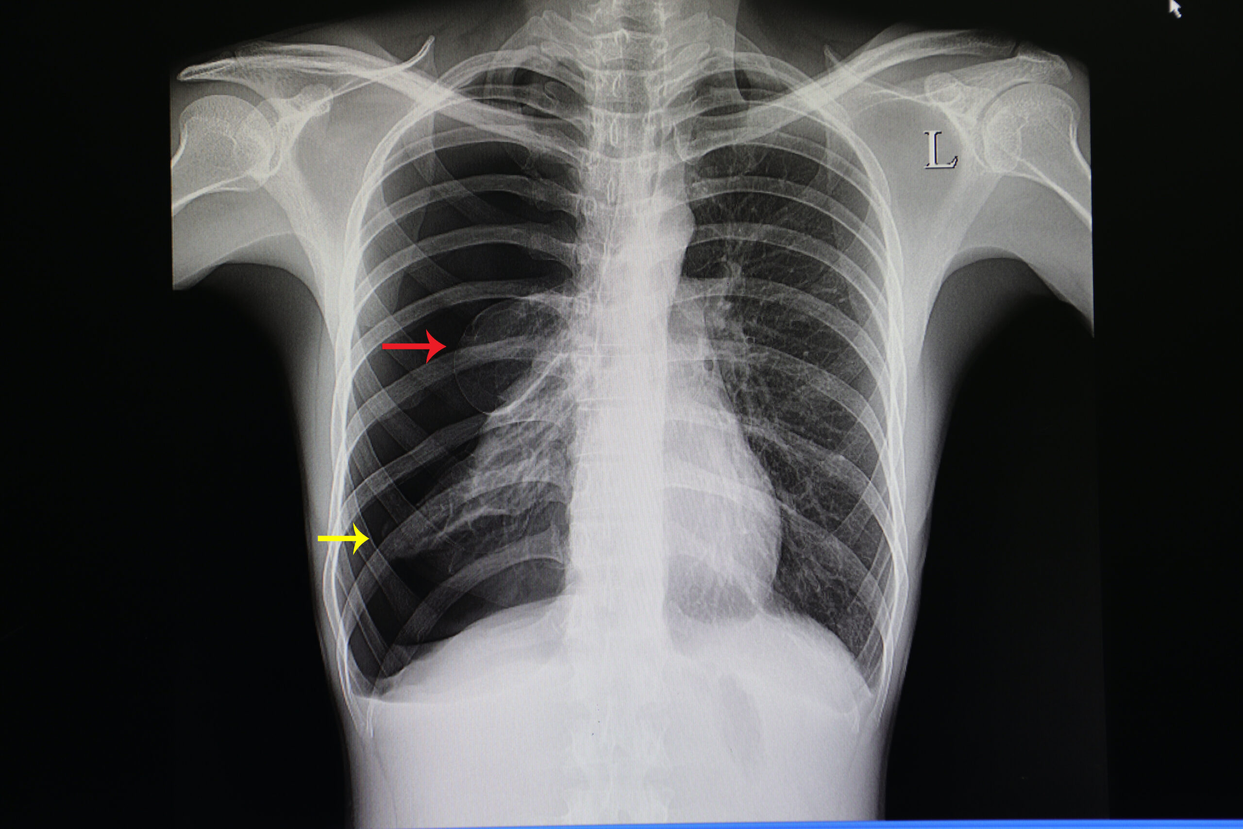
Collapsed Lung X-Ray
Bronchiolitis
Hydration, oral and nasal suctioning, and supplementary oxygen to maintain SpO2 > 94% are supportive interventions for bronchiolitis management. Bronchodilators (nebulized albuterol and epinephrine) and corticosteroids are not routinely recommended. Nebulized medications may aggravate symptoms in some patients. Infants with a history of wheezing or a family history of asthma may benefit from a bronchodilator trial. Nebulized therapy should be discontinued after a trial if there is no significant improvement.25
Asthma26
Asthma is diagnosed as mild, moderate, or severe. Each category has criteria based on breathlessness, ability to talk, level of consciousness, age, the use of accessory muscles for breathing, grading of wheezes, heart rate, and oxygen saturation.
Guide To Assessing Asthma Severity
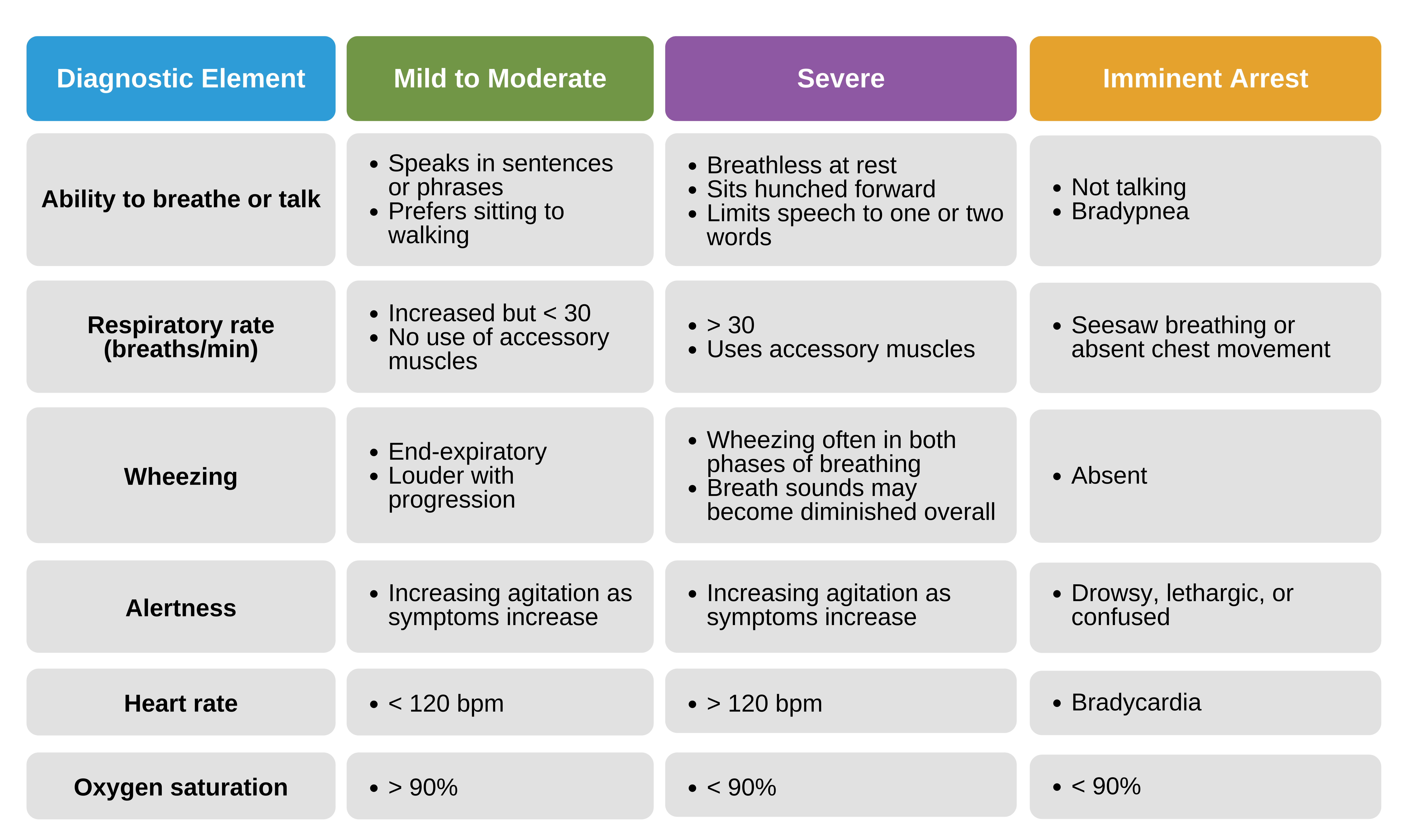
Guide to assessing asthma severity.
Pulsus paradoxus may be present in moderate asthma and is almost certainly detectable in severe asthma. Peak expiratory flow rate (PEFR) measurements are useful if the child’s baseline values are known. PEFR may also be helpful in evaluating response to therapy. PEFR measurements may fatigue a child in moderate to severe distress or worsen bronchospasm.
Treatment for the child with mild to moderate asthma includes:
- Humidified oxygen via a nasal cannula or oxygen mask, titrated for oxygen saturation of at least 94%
- Albuterol via metered-dose inhaler or nebulizer solution
- Oral corticosteroids
Treatment for the child with moderate to severe asthma includes:
- Humidified oxygen via a nasal cannula or oxygen mask, titrated for oxygen saturation of at least 94%
- Albuterol via metered-dose inhaler or nebulizer solution
- Continuous albuterol via nebulization if not improving with intermittent albuterol treatments
- Ipratropium bromide via nebulization
- IV access
- Oral or IV corticosteroids
- Magnesium sulfate IV (BP and HR should be closely monitored)
- High flow nasal cannula or noninvasive positive pressure
- Chest X-ray
- Blood gas analysis
Treatment for the child with severe asthma who presents with severe respiratory distress or imminent respiratory failure should include all the above interventions as well as:
- Oxygen supplementation at high concentrations via a nonrebreather mask
- Continuous albuterol nebulization
- Terbutaline administered subcutaneously or via IV infusion
- Noninvasive ventilation if the child is alert and cooperative
- Intubation with a cuffed ET tube if the child progresses to respiratory failure
Endotracheal intubation may cause respiratory and circulatory complications. Children in status asthmaticus are very difficult to ventilate with positive pressure ventilation and are at high risk for iatrogenic injury such as pneumothorax. Providers should aggressively treat the child with all other interventions and avoid endotracheal intubation whenever possible.
Managing Lung Tissue Disease
Many clinical conditions can cause lung tissue disease, and this section will present the management of the most common conditions, such as pneumonia, cardiogenic pulmonary edema, ARDS, traumatic pulmonary contusion, and allergic, vascular, and inflammatory insults.
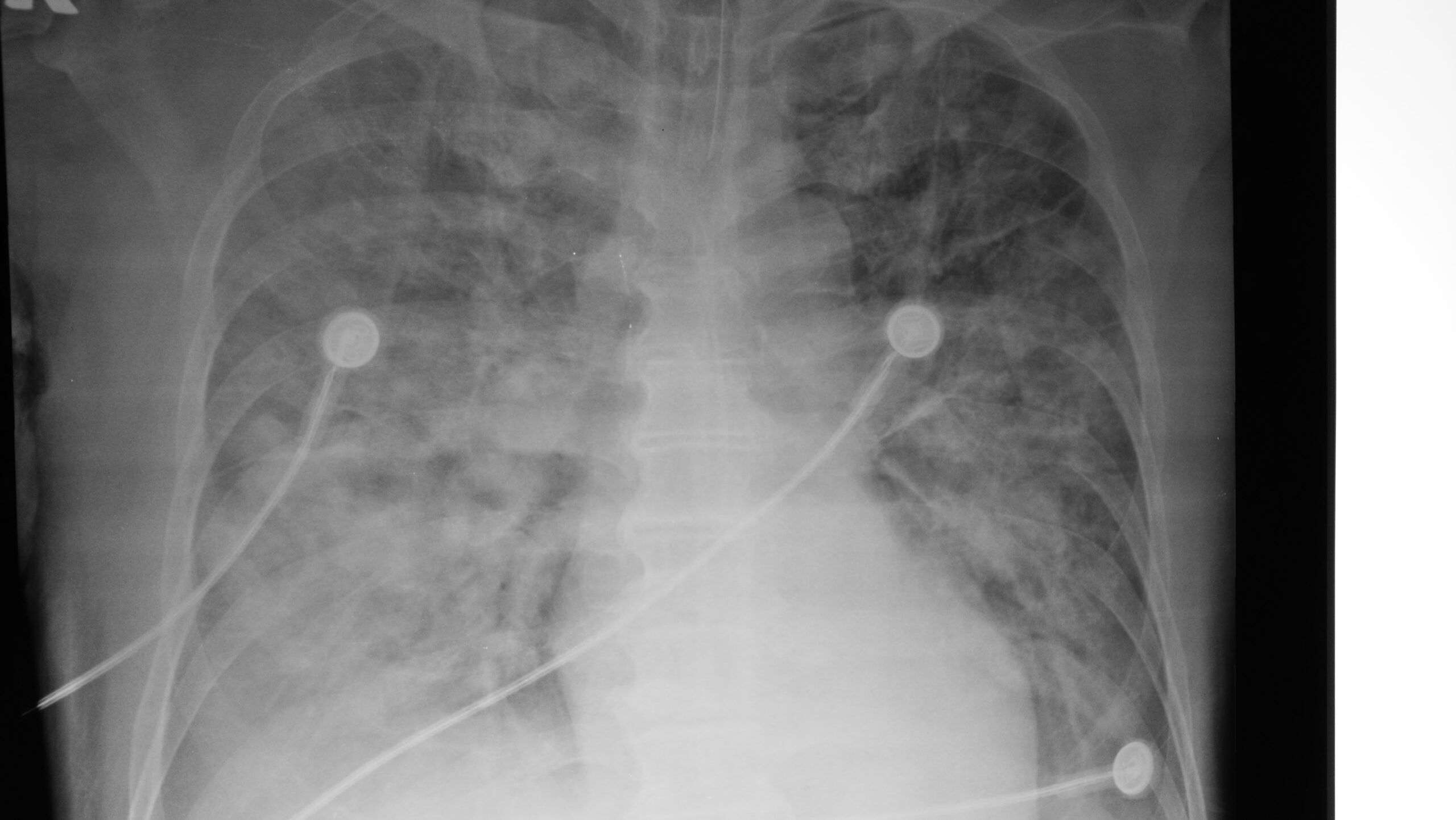
Acute Respiratory Distress Syndrome Lung X-Ray
The general treatment of lung tissue disease includes the initial management of respiratory distress or failure and oxygen supplementation as required. Humidified high-flow nasal cannula oxygen therapy or positive pressure oxygen administration via continuous positive airway pressure (CPAP) noninvasive ventilation, or mechanical ventilation may be beneficial.
Related Video – One Quick Question: How Does CPAP Work?
Infectious Pneumonia
Infectious pneumonia can be due to a viral, bacterial, or fungal infection causing inflammation of the alveoli. Diagnostic procedures such as complete blood count, blood culture and sensitivity studies, viral studies, sputum Gram stain, arterial blood gas, and chest X-ray help clinicians determine the etiology and provide the appropriate treatments.
Appropriate treatment is determined once the provider identifies the offending agent. For bacterial infections, the child should be treated with the most suitable antibiotic based on culture and sensitivity results if available. If there are signs of bronchoconstriction, albuterol via metered-dose inhaler or nebulization should be ordered. In severe cases, noninvasive positive-pressure ventilation may be an option if tolerated. If the patient develops severe respiratory distress or respiratory failure, the team should be prepared for endotracheal intubation.
Chemical Pneumonitis
Aspirating or inhaling toxic vapors, gases, or liquids can cause chemical pneumonitis. Chemical pneumonitis may lead to noncardiogenic pulmonary edema, airway swelling, and gas diffusion impairment at the alveolar-capillary interface.
After performing the initial steps of managing respiratory distress or failure, treatment of the child with chemical pneumonitis may include:
- Supplementary oxygen
- Nebulized bronchodilators to treat wheezing
- CPAP or noninvasive ventilation
- Endotracheal intubation if the patient exhibits signs of severe upper airway edema and obstruction with concomitant respiratory distress or failure
Children with severe chemical pneumonitis may require specialized forms of ventilation, such as high-frequency oscillatory ventilation or extracorporeal cardiopulmonary resuscitation (ECMO) The child should be transported to a facility skilled in such interventions.
Aspiration Pneumonitis
Aspiration of stomach acid or oral secretions can cause inflammation and edema to lung tissues. The bacteria from undigested stomach contents can cause pneumonia. Fever is a typical sign of pneumonitis following an aspiration event.
A chest X-ray and complete blood count aid in the diagnosis of aspiration pneumonia. Treatment includes antibiotics and oxygen as needed. In severe cases, CPAP, noninvasive ventilation, or intubation with mechanical ventilation may be required if the patient progresses to respiratory distress or failure.
Cardiogenic Pulmonary Edema
Pulmonary edema is the extravasation of fluid from capillaries into the interstitial space. Cardiac conditions that cause blood to backflow may increase pressure in the pulmonary arteries and pulmonary veins, causing cardiac pulmonary edema.
In children, congenital heart diseases, myocarditis, cardiomyopathies, and cardiac-depressant drugs (beta-adrenergic blockers, tricyclic antidepressants, calcium channel blockers, and chemotherapeutic agents) can cause a depressed cardiac ejection fraction due to poor myocardial function.
To treat the child with pulmonary edema, the team must provide invasive or noninvasive ventilatory support with positive end-expiratory pressure (PEEP) Intubation and invasive ventilation are likely needed for the patient with persistent hypoxemia despite high levels of supplementary oxygen and noninvasive positive end-expiratory pressure.
Using PEEP during mechanical ventilation helps reduce the need for a high oxygen concentration as it helps keep the alveoli patent while fluid is in the interstitium. The typical starting setting for PEEP is 5 cm H2O, and it can be adjusted upwards until the oxygen saturation is 94% and above. However, excessive PEEP can cause lung hyperinflation and affect the venous return to the heart. In this case, cardiac output and oxygen delivery are compromised.
Medical interventions include the use of diuretics to reduce left atrial pressure, inotropic infusions, and afterload reducing agents to restore cardiac function. The clinician also seeks help from an expert pediatric cardiologist for the management of these patients.
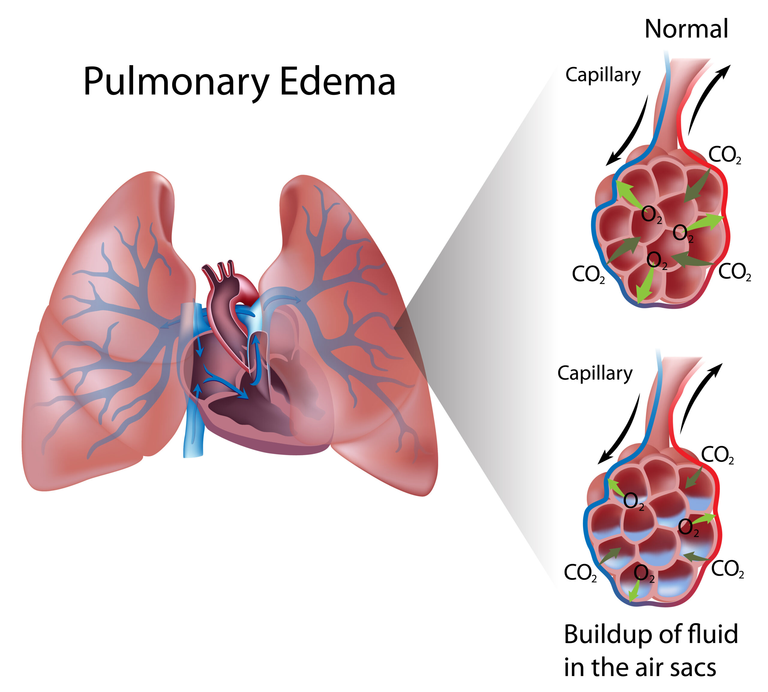
In pulmonary edema, there is a buildup of fluid in the lungs.
Acute Respiratory Distress Syndrome (ARDS)
ARDS is a form of noncardiogenic pulmonary edema following infectious pneumonia, aspiration pneumonia, or a systemic illness such as sepsis or pancreatitis. ARDS can also be triggered by trauma.
These medical conditions cause injury to the alveoli and pulmonary capillaries. The body’s compensatory mechanism for repairing the damage is to recruit inflammatory mediators. As a result, oxygen diffusion into the blood, and to a lesser degree, carbon dioxide diffusion from the blood to the alveoli, are inefficient.
Treatment depends on the diagnosis and usually addresses bacteremia, shock, and respiratory failure.
ARDS is characterized as:
- an acute onset of symptoms within 7 days after the precipitating event or agent
- a PaO2/FiO2 ratio of 300 or less
- an oxygen index of 4 or higher
- formation of new infiltrates on a chest X-ray indicative of parenchymal disease
- no evidence of cardiogenic causes of pulmonary edema
The complexity of this condition necessitates expert consultation.
ARDS is managed by monitoring the heart rate and rhythm, blood pressure, respiratory rate, pulse oximetry, and end-tidal carbon dioxide. The clinician should order an arterial blood gas, a central venous blood gas, and a complete blood count to determine the oxygenation and ventilation status of the patient. The patient likely requires ventilatory support, particularly when there is a worsening of clinical and radiographic findings and persistent hypoxemia despite the delivery of high concentrations of oxygen.
The patient’s hypoxemia must be corrected to prevent the progression of respiratory distress or failure to cardiac arrest. PEEP can be increased to augment oxygen saturation. A lung-protective ventilator strategy should be used during mechanical ventilation. Tidal volumes should be limited to 5–8 mL/kg, and inspiratory plateau pressures should be kept at ≤ 28 cm H2O.27
Managing Disordered Control of Breathing
Disordered control of breathing is usually due to neurologic disorders secondary to increased intracranial pressure (ICP), neuromuscular diseases, CNS infections, traumatic brain injuries, brain tumors, and hydrocephalus. Some conditions and medical interventions can induce disordered control of breathing, such as deep sedation, drug overdose, hyperammonemia, metabolic conditions, and seizures. Clinical findings of increased ICP include unequal pupils not responding to light, disordered breathing, apnea, bradycardia, hypertension, and decerebrate and decorticate positions.
For trauma patients, a patent airway is maintained by positioning the patient and performing the jaw thrust maneuver. Aside from maintaining a patent airway, this maneuver also supports the patient’s spine in case there is spinal involvement from trauma. If there is poor perfusion secondary to poor end-organ function, the provider can administer 20 mL/kg isotonic crystalloid solution or blood products.
Pharmacologic therapy to decrease ICP includes osmotic agents and hypertonic saline solution. After establishing an airway, the team must address the patient’s anxiety and give pain medications to prevent further agitation. Fever should be aggressively treated in these patients to lessen metabolic demands.
The provider must ensure the child receives adequate oxygenation and ventilation. Brief periods of hyperventilation may prevent uncal herniation when there are sudden ICP increases.
Aggressive prophylactic hyperventilation to a PaCO2 of < 30 mm Hg is to be avoided, though. Hyperventilation decreases cardiac output because it hinders the venous return of blood into the heart. Excessive hyperventilation also causes cerebral vasoconstriction, inhibiting blood perfusion and potentially causing cerebral ischemia. Mild hyperventilation is only indicated in the first 48 hours after the injury for acute signs of cerebral herniation. These patients must be monitored for cerebral ischemia.
Key Takeaway
Hyperventilation may reduce intracranial pressure but must be used carefully in pediatric patients.
Increased Intracranial Pressure
Increased ICP disrupts the breathing centers of the brain. Certain conditions such as subarachnoid hemorrhage, tumor growth, meningitis, encephalitis, intracranial abscess, subdural hematoma, and epidural hematoma can cause increased ICP. Patients will present with an irregular respiratory rate and respiratory pattern.
Patients can present with the Cushing triad, which includes a change in respirations, bradycardia, and widening pulse pressure. The triad may herald imminent uncal herniation, which is fatal. The team must seek expert consultation from a neurosurgeon. Specific management depends on the etiology.
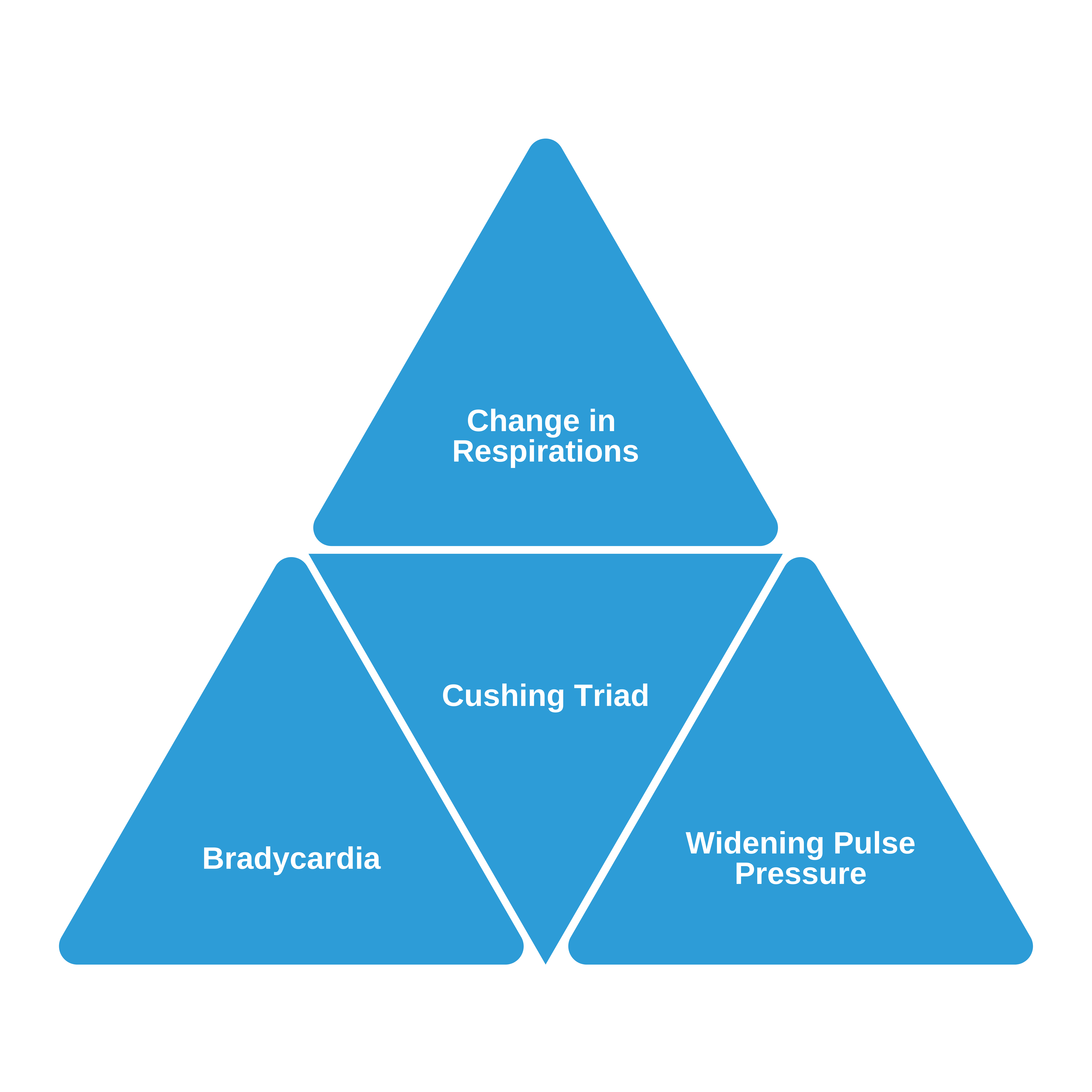
Cushing Triad
Neuromuscular Disease
Chronic progressive neuromuscular disease is potentially fatal when the muscles for respiration are affected. Preliminary symptoms include ineffective coughing and difficulty removing secretions. Due to the damaged muscles, certain complications arise, such as atelectasis, aspiration pneumonitis, pneumonia, restrictive lung disease, and, eventually, respiratory failure. If the patient has an advanced neuromuscular disorder, they may require long-term noninvasive ventilation support.
Succinylcholine, a drug commonly used for intubation, should not be administered to a child with neuromuscular disease. This medication may trigger a life-threatening condition such as hyperkalemia or malignant hyperthermia. Antibiotics in the aminoglycoside family are also known to cause neuromuscular blockade and should be avoided in a child with a neuromuscular disorder.
Drug Overdose and Poisoning
CNS depression from narcotic use or prescription drug overdose may eventually lead to respiratory distress or failure. Sometimes, these drugs cause the temporary loss of function of respiratory muscles and loss of consciousness, leading to upper airway obstruction by the tongue.
After performing the initial management of respiratory distress and failure, the team seeks expert consultation with a toxicologist. If the child has vomited, the team may need to suction the gastric contents to prevent aspiration. If the offending agent is identified and has an antidote, the provider administers the antidote as recommended. For example, a child with an opioid overdose requires the antidote naloxone. Standard BLS and PALS measures are never delayed while waiting on an antidote.
After the initial treatment, other diagnostic assessment tools to guide the next interventions for the patient include an arterial blood gas, ECG, chest X-ray, electrolytes, blood glucose, serum osmolality, and drug screening tests.
When responding to a child with a suspected opioid overdose and resultant respiratory arrest, rescue breathing should be administered until spontaneous breathing begins. The child in respiratory arrest should receive intranasal or intramuscular naloxone. However, standard PALS measures (CPR) should be the priority for the child in suspected cardiac arrest.
25Ralston SL, Lieberthal AS, Meissner HC, et al. Clinical practice guideline: the diagnosis, management, and prevention of bronchiolitis. Pediatrics. 2014;134(5):e1474–e1502.
https://pediatrics.aappublications.org/content/134/5/e1474.short
26Expert Panel Working Group of the National Heart, Lung, and Blood Institute (NHLBI) administered and coordinated National Asthma Education and Prevention Program Coordinating Committee (NAEPPCC), Cloutier MM, Baptist AP, et al. 2020 focused updates to the asthma management guidelines: a report from the National Asthma Education and Prevention Program Coordinating Committee Expert Panel Working Group. J Allergy Clin Immunol. 2020;146(6):1217–1270.
27The Pediatric Acute Lung Injury Consensus Conference Group. Pediatric acute respiratory distress syndrome: consensus recommendations from the pediatric acute lung injury consensus conference. Pediatr Crit Care Med. 2015;16(5):428–439.