Cardiac Arrest in Infants and Children
The survival rate of the pediatric patient depends on where the arrest has taken place (in-hospital vs. out of hospital) and the presenting rhythm of the arrested child. The largest proportion of pediatric cardiac arrests occur at home or in other non-public places. However, survival rates are higher in patients who have an in-hospital cardiac arrest (IHCA).
Nonshockable rhythms are the most frequent in pediatric patients, seen in 82% of cardiac arrests. But survival rates are higher with shockable cardiac arrest rhythms—20% compared to 5% with patients presenting with nonshockable rhythms in one population-based study.14
Key Takeaway
Early recognition and management of respiratory distress, respiratory failure, and shock are critical to prevent progression to cardiac arrest.
The highest survival rates occur when CPR is performed in patients with significant bradycardia and poor perfusion before pulseless arrest develops. However, there are poor survival outcomes to hospital discharge or significant neurologic impairments in many of these patients.15 Recognition and intervention prior to arrest are critical to short- and long-term outcomes.
Definition of Cardiac Arrest
Cardiac arrest is the absence of blood circulation due to the cessation of mechanical activity of the heart. Patients in cardiac arrest have absent pulses. Pediatric patients are unresponsive with abnormal breathing, agonal gasps, and cessation of breathing. Cerebral hypoxia leads to loss of consciousness. The resulting multiorgan ischemia leads to death if the team does not intervene quickly.
Cardiac Arrest Pathways
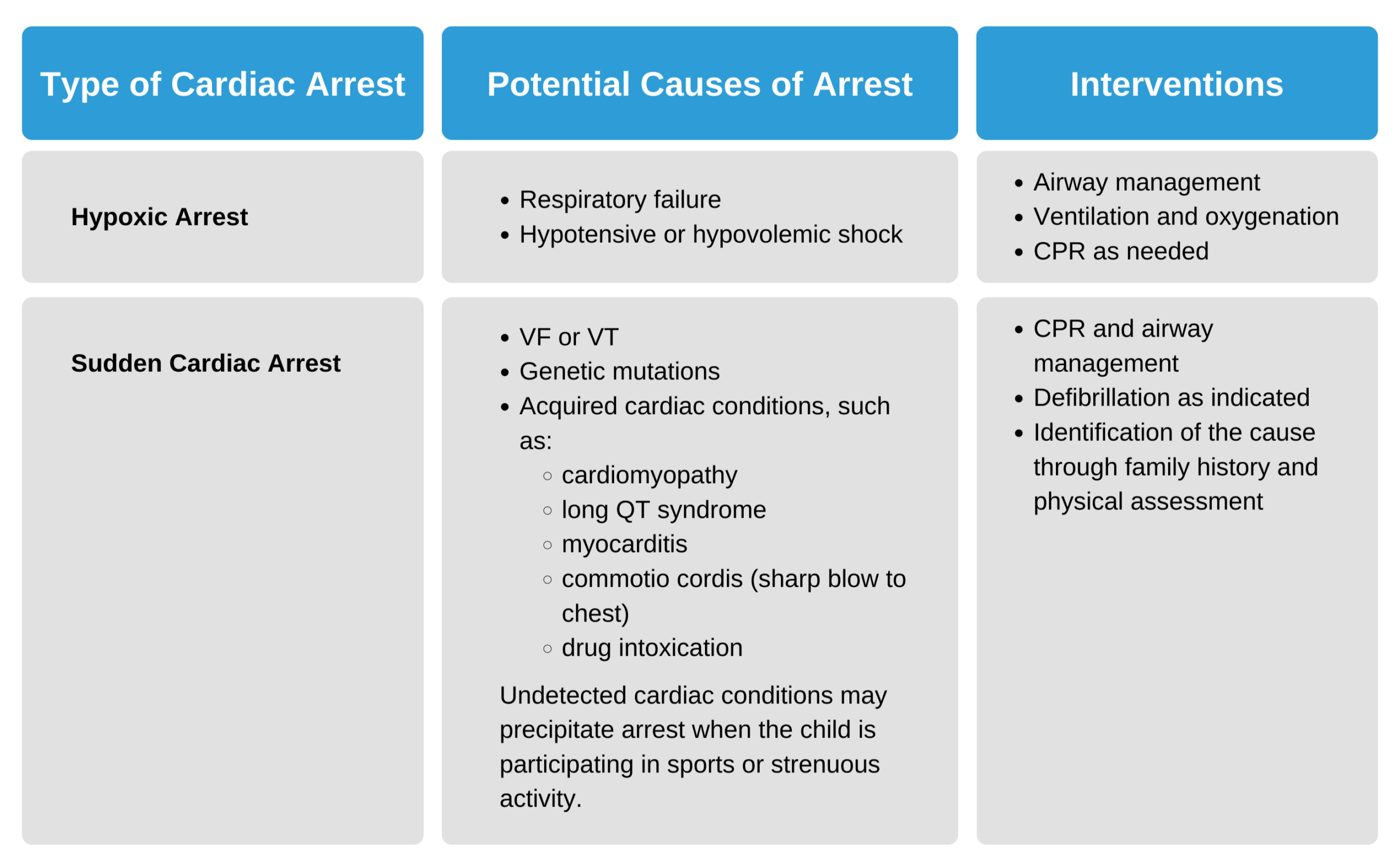
Hypoxic arrest and sudden cardiac arrest are the two pathways for cardiac arrest.

Cardiac Arrest Pathways
Causes of Cardiac Arrest
Out-of-hospital cardiac arrest in infants and children mostly occurs at home. Sudden unexpected infant death (SUID) is a leading cause of death in infants younger than 12 months of age in the United States.16 Infant and parent co-sleeping increases the risk of death by asphyxiation Interventions such as placing the infant sleep on their back or giving babies their own space to sleep have decreased the incidence of cardiac arrest in the infant population.17
The majority of cardiac arrests in children result from a primary respiratory compromise.18 However, arrhythmias are the most frequent cause of cardiac arrest in adults.
Another cause of cardiac arrest in children is trauma. In traumatic cardiac arrest, patients may have airway compromise, traumatic tension pneumothorax, hemorrhagic shock, or brain injury.
Children at risk of cardiac arrest have inadequate oxygenation, ventilation, and perfusion. The PALS provider must detect and treat respiratory distress, respiratory failure, and shock before the child progresses into cardiopulmonary failure and cardiac arrest.
For the child in cardiac arrest, immediate high-quality CPR is necessary. The PALS provider must look for reversible causes of cardiac arrest so that the team can perform the required interventions to reverse the arrest.
The mnemonic Hs and Ts is an essential tool for evaluating the reversible causes of cardiac arrest and quickly intervening.
- Hypovolemia
- Hypoxia
- Hydrogen ion (acidosis)
- Hypoglycemia
- Hypo-/Hyperkalemia
- Hypothermia
- Tension pneumothorax
- Tamponade, cardiac
- Toxins
- Thrombosis, pulmonary
- Thrombosis, coronary
- Trauma
Related Video – Hs and Ts – Hypovolemia
Related Video – Hs and Ts – Hypoxia
Related Video – Hs and Ts – Hydrogen Ions
Related Video – Hs and Ts – Hypo-Hyper Kalemia
Related Video – Hs and Ts – Tension Pneumothorax
Related Video – Hs and Ts – Pulmonary Embolism
Related Video – Hs and Ts – Cardiac Tamponade
Related Video – Hs and Ts – Hypothermia
Related Video – Hs and Ts – Thrombosis Coronary
Related Video – Hs and Ts – Toxins
Recognition of Cardiac Arrest
A pediatric patient in cardiac arrest presents as unresponsive, apneic, or with abnormal breathing and without a detectable pulse. These clinical findings should be enough to recognize cardiac arrest. In the hospital setting, a cardiac monitor may give additional information about an arrest rhythm. Cardiac monitoring is also an excellent tool for continuously assessing the child. The child should be connected to a cardiac monitor as quickly as possible in the hospital setting.

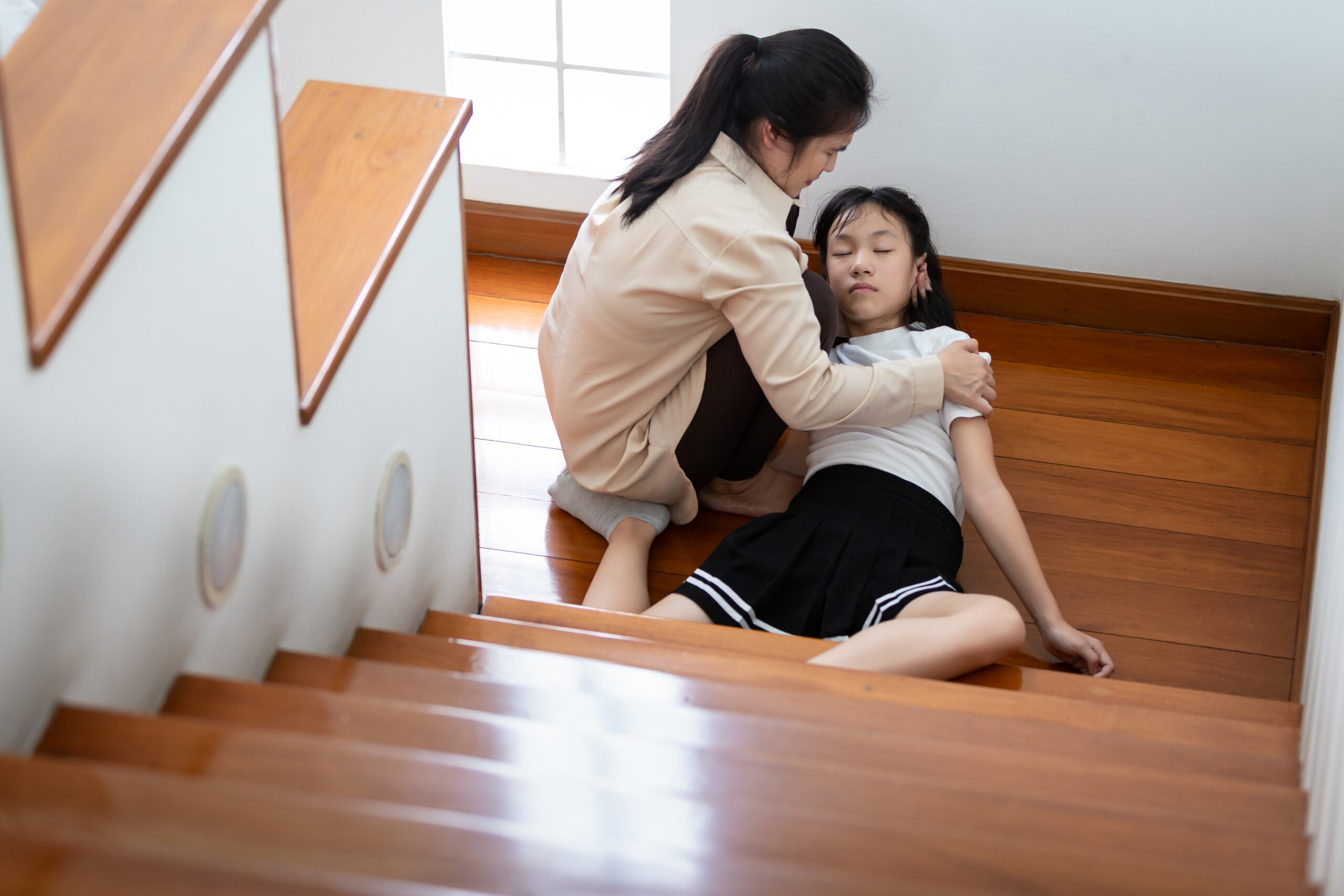
Unresponsive Child
When performing the primary assessment on a critically ill child, assessing responsiveness, breathing, and checking for a pulse should not take more than 10 seconds.
Key Takeaway
Start high-quality CPR if a pulse is not felt within 10 seconds!
Palpating for pulses in infants and children can be difficult. In an infant, the brachial pulse is palpated, while the femoral or carotid pulses are palpated in children. Even experienced providers may have difficulty palpating a pulse in an infant or child. The PALS provider should conclude that the patient is in cardiac arrest and proceed with high-quality CPR if a pulse is not detected within 10 seconds.
Cardiac arrest is associated with four rhythms categorized into shockable and nonshockable rhythms.
Key Takeaway
Cardiac arrest rhythms in children include:
Shockable rhythms:
VF and pVT
Nonshockable rhythms:
asystole and PEA
Shockable rhythms require defibrillation to convert them into a normal rhythm and restore cardiac function. Ventricular fibrillation and pulseless ventricular tachycardia are shockable rhythms.
Nonshockable rhythms cannot be treated with defibrillation but require immediate chest compressions and medications. Asystole and pulseless electrical activity (PEA) are nonshockable rhythms.
Related Video – One Quick Question: Should You Choose CPR or Defibrillation?
Ventricular Fibrillation
Related Video – ECG Rhythm Review – Ventricular Fibrillation
Ventricular fibrillation (VF) has no organized rhythm or coordinated myocardial contractions. VF’s electrical activity is chaotic, which represents a quivering heart incapable of producing significant cardiac output.
VF can be graded from coarse to fine, depending on the amplitude of the deflections. Coarse VF may represent a relatively new onset, while fine VF may occur when the rhythm has been present for a significant amount of time without medical intervention. Fine VF deteriorates to asystole if left untreated.
Patients in VF have no pulse because there is no substantial cardiac output. Without intervention, patients in ventricular tachycardia with a pulse or in pulseless ventricular tachycardia may develop VF. VF has been reported in up to 27% of IHCA pediatric patients at some time while performing resuscitation.19
Cardiac anomalies, such as cardiac ion channelopathies, can produce VF. Other causes include a traumatic blow to the chest (commotio cordis). Better survival rates are observed in VF, VT, and pVT as compared with asystole and PEA.
Pulseless Ventricular Tachycardia (pVT)
Related Video – ECG Rhythm Review – Ventricular Tachycardia
Ventricular tachycardia can present with or without a pulse. Pulseless ventricular tachycardia (pVT) is the arrest rhythm requiring defibrillation and CPR. Ventricular tachycardia with a pulse often deteriorates to pVT or VF if not treated rapidly. Ventricular tachycardia is an organized rhythm, in contrast with VF, where the rhythm is disorganized.
pVT has a wide QRS complex and is usually present only briefly, degrading quickly into VF. In rare cases, patients may present in VT with a pulse and remain stable for quite some time. When a child is in VT, the cardiac monitor displays the rhythm, but it cannot determine whether the patient has a pulse. The monitor is not a substitute for clinical assessment skills. Where a child in stable VT may be treated with medication, the team should treat the child in pVT as if they are in VF.
In monomorphic VT, the QRS complexes all have the same amplitude. Their amplitude varies in polymorphic VT. When the polymorphic VT rhythm twists along the isoelectric line, it is known as torsades de pointes Torsades de pointes is a specific arrhythmia that is treated differently than other cases of polymorphic VT as it has a few causes, including congenital long QT syndrome, hypomagnesemia, and drug toxicity.
Related Video – ECG Rhythm Review – Polymorphic Ventricular Tachycardia (Torsades de Pointes)
Asystole
Asystole represents a nonfunctioning myocardium devoid of any electrical activity. It appears as a flat line on an ECG. When the PALS provider sees that a patient is in asystole, they must check for a pulse, make sure that all ECG leads are connected, and determine that the ECG machine is not faulty. They should also increase the gain or amplitude on the ECG monitor to ensure the patient is genuinely in asystole. A patient that has a pulse and is breathing is not in asystole!
Related Video – ECG Rhythm Review – Asystole
Pulseless Electrical Activity (PEA)
Related Video – What is PEA?
PEA can present as any organized rhythm on the ECG monitor, but the patient will not have a palpable pulse. The monitor may show a slow, normal, or fast rhythm. PEA can also exhibit low or high amplitude T waves, prolonged PR and QT intervals, atrioventricular dissociation, complete heart block, or ventricular complexes without P waves.
A common cause of PEA in life-threatening situations is hypovolemia. If PEA is left untreated, it will convert into asystole. The three rhythms that cannot mimic PEA are VF, pVT, and asystole.
14 Atkins DL, Everson-Stewart S, Sears GK, et al. Epidemiology and outcomes from out-of-hospital cardiac arrest in children: the Resuscitation Outcomes Consortium Epistry-Cardiac Arrest. Circulation. 2009;119(11):1484–1491.
15 Slomine BS, Silverstein FS, Christensen JR, et al. Neuropsychological outcomes of children 1 year after pediatric cardiac arrest: secondary analysis of 2 randomized clinical trials. JAMA Neurol. 2018;75(12):1502–1510.
16 Data and statistics: fast facts. Centers for Disease Control and Prevention website. Accessed October 21, 2021.
17 Task Force on Sudden Infant Death Syndrome. SIDS and other sleep-related infant deaths: updated 2016 recommendations for a safe infant sleeping environment. Pediatrics. 2016;138(5):e20162938.
https://pediatrics.aappublications.org/content/138/5/e20162938
18 Duff JP, Topjian AA, Berg MD, et al. 2019 American Heart Association focused update on pediatric advanced life support: an update to the American Heart Association guidelines for cardiopulmonary resuscitation and emergency cardiovascular care. Pediatrics. 2020;145(1):e20191361.
https://pediatrics.aappublications.org/content/145/1/e20191361.long
19 Samson RA, Nadkarni VM, Meaney PA, et al. Outcomes of in-hospital ventricular fibrillation in children. N Engl J Med. 2006:354(22):2338–2339.