Inserting the ET Tube: Tips for Calculating Depth
This guide covers the essential formulas and techniques for estimating endotracheal (ET) tube insertion depth. Designed for healthcare providers and emergency responders, this article will teach you how to quickly calculate the correct depth, establish a consistent reference point, and properly confirm placement to minimize complications during intubation.
ACLS Certification Association videos have been peer-reviewed for medical accuracy by the ACA medical review board.
Article at a Glance
The Quick Answer: To quickly estimate the proper endotracheal tube depth for an adult, multiply the tube’s internal diameter by 3 and secure that measurement at the patient’s teeth.
- Before insertion, the provider must estimate the tube’s depth.
- The markings on the ET tube determine the distance from the end of the tube.
- The provider should multiply the ET tube size by 3.
- The ET tube is inserted and secured to that calculated number at the patient’s teeth (specifically the upper incisors) to ensure consistent documentation.
- Tube placement must be evaluated after insertion using multiple modalities, including auscultation and continuous waveform capnography (CO₂ measurement).
Calculating Tube Depth
Accurately calculating the endotracheal tube depth prevents complications like right mainstem intubation (a dangerous form of endobronchial intubation). The calculation relies on understanding the tube’s size and markings, though it serves as a rough estimate and may vary based on individual patient factors.
Step 1: Identify ET Tube Size (Inner Diameter)
First, look at the tube to identify the size, called the inner diameter (ID). In this example, we will reference a 7.0 ET tube.

The size of the tube is called the inner diameter. In this example, the size is 7.0.
Step 2: Understand Tube Markings
There are also numbered markings along the side of the endotracheal tube. These markings measure the distance in centimeters from the distal end of the tube (the tube tip) up to that particular mark.

Notice that there are numbered markings along the side of the endotracheal tube.
Step 3: Calculate Depth (Size × 3)
- Take the ET tube size and multiply it by 3.
- For a 7.0 ET tube, you would multiply 7 times 3 to equal 21.
- When inserting the endotracheal tube into the patient and passing it through the vocal cords, the 21 cm mark should sit at the patient’s teeth (specifically the upper incisors).
Read: General Stroke Care

Note the marking number at the patient’s teeth. The number of the marking at the teeth should be approximately the ET tube size multiplied by three.
Alternative Method: Height-Based Formula
Because the “Size × 3” rule is a rough estimate, an alternative approach commonly used by clinicians is to estimate depth based on the patient’s height. For example, a standard height-based formula is: Depth (cm) = (Height in cm / 10) + 5. For more precision, you can consult external clinical guidelines on height-based intubation formulas.
Confirming Placement
Of course, you will still assess for lung sounds when evaluating the initial et tube placement depth. However, confirmation must expand beyond auscultation. Providers should utilize continuous CO₂ measurement (capnography), obtain a chest radiograph (X-ray) for prolonged intubation, and can use ultrasound as a quick adjunct to ensure proper tube position.
Common Mistakes to Avoid
- Misreading the numbered markings on the side of the tube.
- Using an inconsistent reference point (e.g., measuring at the lips instead of the teeth/upper incisors).
- Over-relying on a single rule of thumb without confirming via clinical assessment and capnography.
Related Video – One Quick Question: How do You Fix ET Tube Adaptor Issues??
Summary & Next Steps
Correct tube depth is a critical part of successful endotracheal intubation, since improper placement can lead to poor delivery of tidal volume, inadequate overall ventilation, or right mainstem bronchus intubation. Follow this post-intubation checklist to ensure a secure airway:
- Calculate: Multiply the ET tube’s internal diameter by 3 (e.g., 7.0 × 3 = 21 cm).
- Align: Secure the calculated marking number directly at the patient’s upper teeth (incisors).
- Confirm: Validate placement immediately using auscultation (sight and sound), CO₂ capnography, and a chest radiograph.
More Free Resources to Keep You at Your Best
Editorial Note
ACLS Certification Association (ACA) uses only high-quality medical resources and peer-reviewed studies to support the facts within our articles. Explore our editorial process to learn how our content reflects clinical accuracy and the latest best practices in medicine. As an ACA Authorized Training Center, all content is reviewed for medical accuracy by the ACA Medical Review Board.
More to Learn
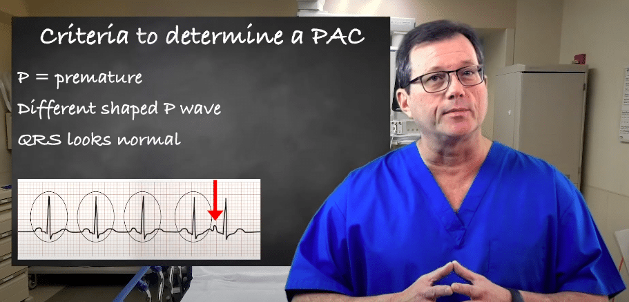
Explore premature atrial contractions (PACs) and the three determinants used to identify them. Our article will enhance your understanding of cardiac arrhythmias.
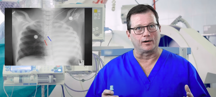
Learn how to correct right mainstem intubation and achieve the target range for endotracheal (ET) tube placement. Our article helps enhance patient safety and airway management.