
Approach to ECG Interpretation – The P Wave
The P wave is upright, rounded, and uniform. It usually occurs before the QRS complex, though in certain circumstances, it may occur during or after the QRS complex. The QRS complex is the most easily visualized marking on the ECG strip due to its size.
When an impulse originates somewhere other than the SA node, the P wave morphology changes, and it may sometimes have a downward deflection. Occasionally, a P wave may be difficult to identify because it is superimposed on another ECG waveform. Figure 2.11 illustrates first-degree AV delay, traditionally called first-degree AV block. In this condition, every P wave is conducted to a QRS complex, but the PR interval is prolonged beyond 0.20 second. When the PR interval is markedly prolonged, atrial depolarization may occur during the preceding ventricular repolarization, causing the P wave to overlap the preceding T wave. The P wave represents atrial depolarization, the QRS complex represents ventricular depolarization, and the T wave represents ventricular repolarization. The relatively small P wave may be difficult to distinguish when it is superimposed on the larger T wave. The P wave has not disappeared; it is visually obscured by the larger T wave. This is commonly described as a hidden or buried P wave.
Figure 2.11 First-Degree Atrioventricular Block
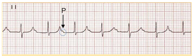
First-Degree AV Block ECG
Possible causes of a buried P wave:
- Increased vagal tone
- Athletic training
- Inferior myocardial infarction
- Mitral valve surgery
- Myocarditis from Lyme disease
- AV nodal-blocking drugs
The P wave is the most reliable wave to identify on the ECG strip. All leads should be examined for P waves. The absence of clearly identifiable P waves may indicate atrial fibrillation or another arrhythmia.
The relationship between P waves and the QRS complex should also be established. The number of P waves should equal the number of QRS complexes.
- If there are more QRS complexes than P waves, then the rhythm is an accelerated ventricular or junctional rhythm.
- If there are more P waves than QRS complexes, then some type of AV block is present.
- P waves that occur after each QRS complex may indicate a junctional rhythm, a ventricular rhythm with retrograde AV conduction, an AV nodal re-entrant rhythm, or an AV-reciprocating tachycardia.