Electrophysiology – The Conduction System
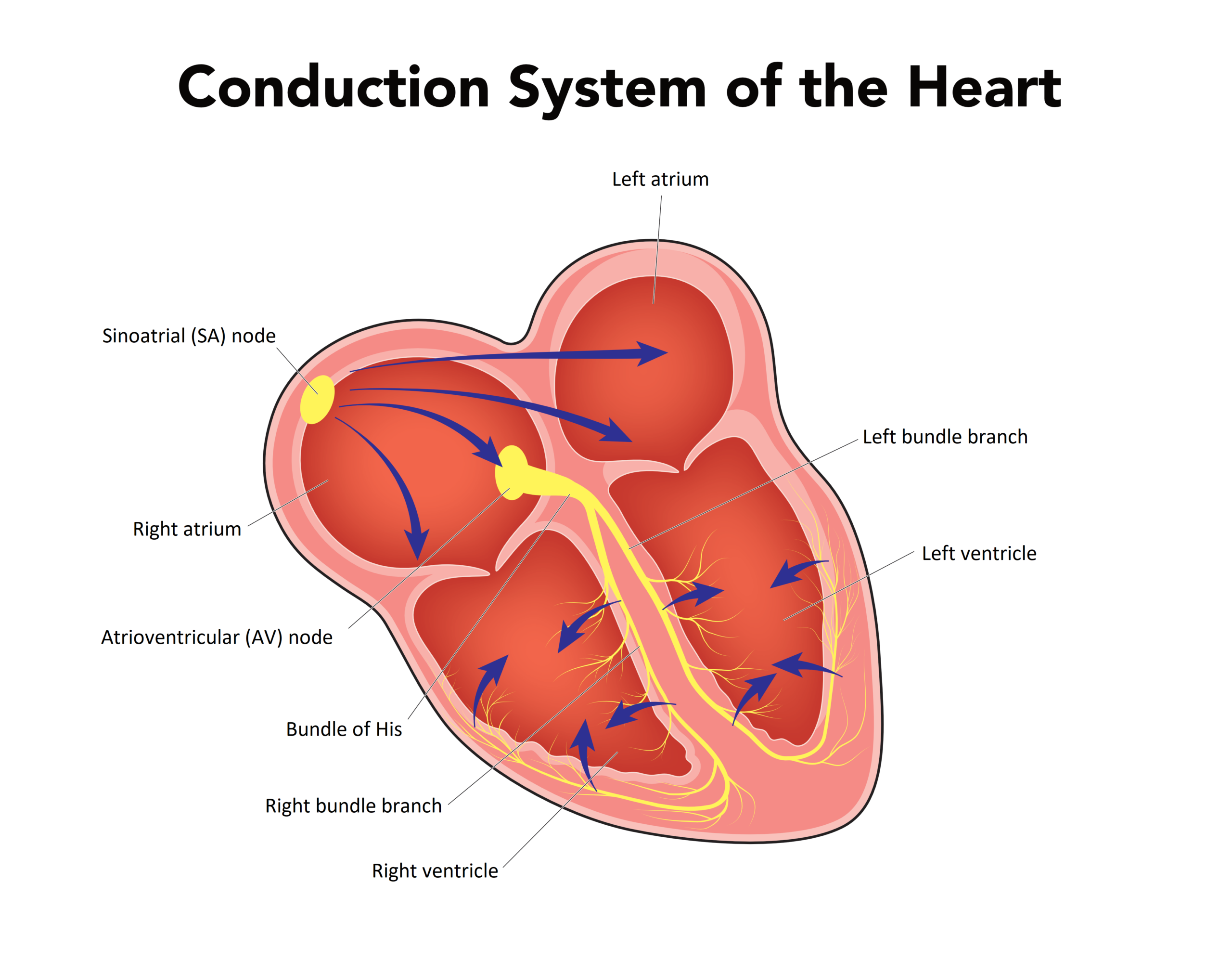
The conduction system of the heart is arranged in a system of pathways, much like a circuit board. The arrangement efficiently transfers electrical energy to the cardiac muscle cells for contraction (see Figure 1.4). This system contains the sinoatrial node (SA node), in which the first electrical impulse is generated by the node’s pacemaker cells. The SA node is in the right atrium near the superior vena cava. It is composed of a parasympathetic nerve (cranial nerve X, the vagus nerve) and sympathetic nerves (T1–T4 spinal nerves).
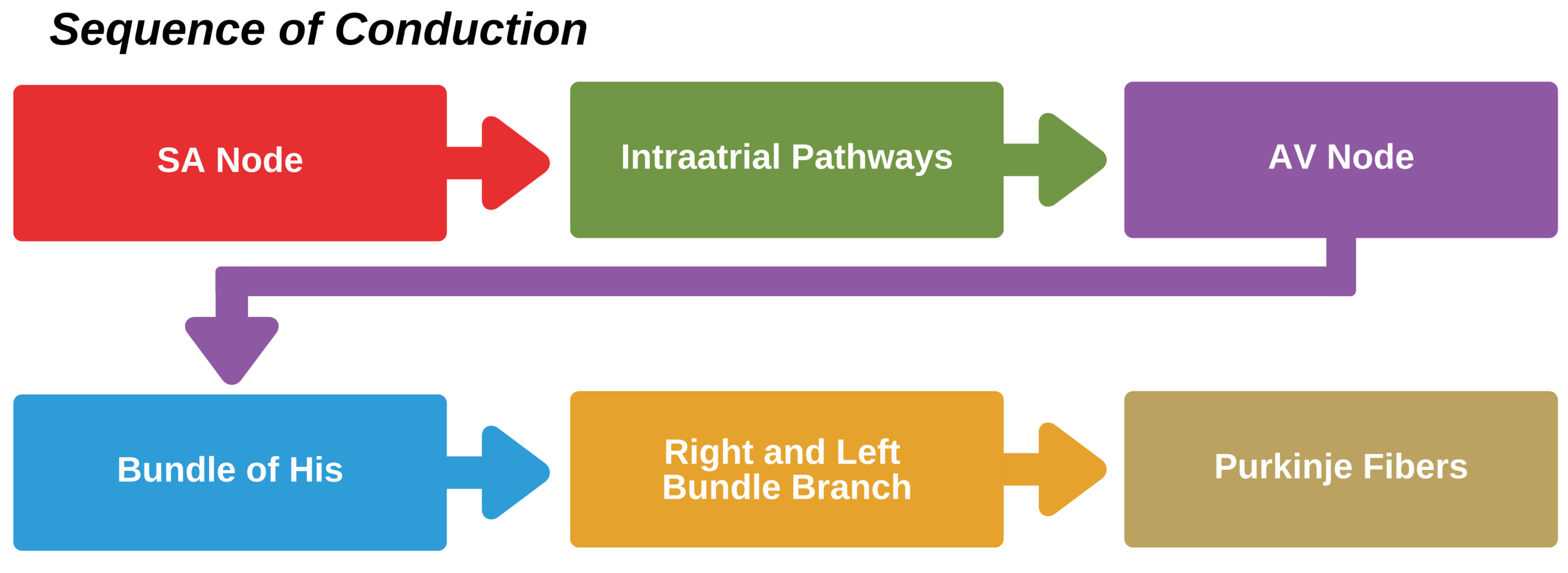
The impulses generated from the SA node travel through the right and the left atria to the atrioventricular node (AV node). After the AV node, the impulse travels to the bundle of His and then to the right and left bundle branches. At the ends of these bundle branches are terminal branches made up of small fibers, known as the Purkinje fibers. These fibers are connected to the muscle cells and stimulate contraction (see Figure 1.5).
Figure 1.4. The Heart’s Conduction System

Heart’s Conduction System
Figure 1.5. The Sequence of Conduction

The Sequence of Conduction Infographic