Hyperosmolar Hyperglycemic Syndrome
Hyperosmolar hyperglycemic syndrome (HHS) is a life-threatening diabetic emergency marked by extremely high blood sugar, high serum osmolality, and minimal or absent ketoacidosis. This article explains the definition, causes, symptoms, diagnosis, and treatment priorities for HHS, including fluids first, electrolyte correction, insulin when safe, and treatment of the precipitating cause.
ACLS Certification Association videos have been peer-reviewed for medical accuracy by the ACA medical review board.
Table of Contents
- What is Hyperosmolar Hyperglycemic Syndrome (HHS)?
- What is the difference between DKA and HHS?
- What causes hyperosmolar hyperglycemic state?
- Etiology and Pathophysiology
- Insulin Resistance in Type 2 Diabetes
- Signs and Symptoms of Hyperglycemia
- Diagnosis and Tests
- Management of Hyperosmolar Hyperglycemic Syndrome
- The Dangers of Hypovolemia
- Minimal Ketones in Hyperosmolar Hyperglycemic Syndrome
- Watch out for Hypokalemia
- Summary
Article at a Glance
- HHS is a medical emergency involving severe hyperglycemia, hyperosmolality, and severe dehydration with minimal or absent ketoacidosis.
- HHS is most often associated with type 2 diabetes, but overlap with DKA can occur, so clinicians should avoid strict type 1 vs. type 2 assumptions.
- Fluids come first. Early management focuses on restoring circulating volume, checking electrolytes, replacing potassium as needed, starting insulin when safe, treating the trigger, and monitoring the patient closely.
What is Hyperosmolar Hyperglycemic Syndrome (HHS)?
Hyperosmolar hyperglycemic syndrome (HHS) is a serious complication of diabetes in which glucose rises very high, water is lost through osmotic diuresis, and the blood becomes highly concentrated. The older term hyperosmolar hyperglycemic nonketotic syndrome is still sometimes used, but HHS is now more commonly described by its current clinical name.
Unlike classic DKA, HHS usually has enough circulating insulin to limit major ketone production, but not enough insulin activity to control hyperglycemia. This is why HHS can produce extreme hyperglycemia without the same degree of ketoacidosis usually seen in DKA.
What is the difference between DKA and HHS?
The main difference between HHS and DKA is the degree of ketoacidosis. DKA is driven by significant insulin deficiency, ketone production, and metabolic acidosis, while HHS usually involves enough residual insulin to limit ketone formation but not enough to prevent extreme hyperglycemia and hyperosmolality.
Some patients have overlapping features of DKA and HHS. For SEO and terminology coverage, these overlap patterns may be searched using phrases such as diabetic ketoacidosis hyperglycemic crisis, diabetic ketoacidosis hyperosmolar overlap, ketoacidosis hyperglycemic hyperosmolar overlap, or ketoacidosis hyperosmolar hyperglycemic overlap, but the clinical priority is to evaluate glucose, osmolality, ketones, pH, bicarbonate, potassium, and mental status.
What is Diabetic Ketoacidosis (DKA)?
This video reviews DKA, including how insulin deficiency leads to ketone production, acidosis, and emergency treatment needs. It fits here because DKA is the key comparison point for understanding how HHS differs from ketoacidosis.
What causes hyperosmolar hyperglycemic state?
HHS develops when insulin activity is not enough to control glucose and ongoing osmotic diuresis causes a large fluid and electrolyte deficit. As glucose rises, the kidneys try to clear excess glucose through the urine, which can worsen volume depletion and electrolyte imbalance.
The most common trigger category is infection, especially pneumonia, urinary tract infection (UTI), and sepsis. Other triggers include missed or inadequate diabetes medication, newly recognized diabetes, stroke, myocardial infarction, surgery, trauma, corticosteroids, diuretics, and reduced oral fluid intake during illness.
Risk factors
- Older age, frailty, or limited ability to recognize thirst
- Known or undiagnosed type 2 diabetes mellitus
- Acute infection such as pneumonia, UTI, or sepsis
- Comorbid illness such as kidney disease, heart disease, or stroke
- Medication contributors such as corticosteroids or diuretics
- Delayed recognition because thirst, fatigue, weakness, and frequent urination may develop gradually
Etiology and Pathophysiology
HHS is a severe hyperglycemic emergency that occurs when relative insulin deficiency and insulin resistance combine with increased counterregulatory hormones. DKA is more common in type 1 diabetes and HHS is more common in type 2 diabetes, but mixed presentations can occur.
During acute illness or physiologic stress, hormones such as cortisol, epinephrine, norepinephrine, glucagon, and growth hormone increase glucose production and oppose the action of insulin. In HHS, residual insulin activity usually limits major ketone production, but it is not enough to prevent severe hyperglycemia.
As glucose rises, osmotic diuresis causes fluid and electrolyte loss. If the patient cannot replace those losses, dehydration worsens, serum osmolality rises, kidney perfusion declines, and glucose clearance becomes even more impaired.
Important precipitating factors include infection, especially pneumonia, UTI, and sepsis, as well as missed diabetes therapy, newly recognized diabetes, myocardial infarction, stroke, surgery, trauma, corticosteroids, diuretics, and other illnesses that increase physiologic stress.
The neurologic symptoms of HHS are closely tied to hyperosmolality and dehydration. As osmolality rises, patients may develop confusion, lethargy, focal neurologic deficits, seizures, or coma.
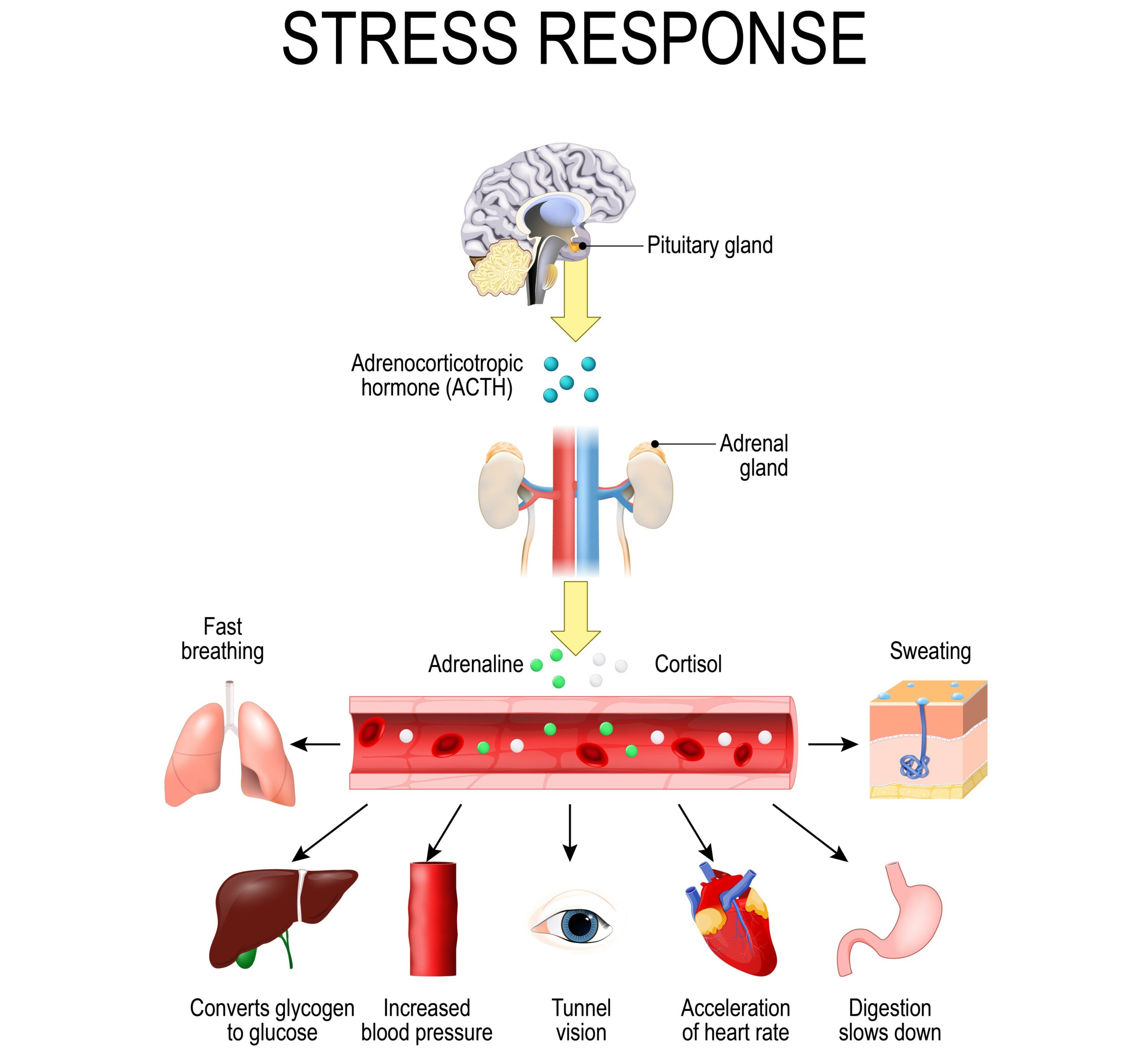
The stress response involves the release of cortisol, epinephrine (adrenaline), and norepinephrine.
Insulin Resistance in Type 2 Diabetes
Insulin resistance is one reason HHS is more often associated with type 2 diabetes. In insulin resistance, the body still produces insulin, but muscle, liver, and fat cells do not respond to it effectively, allowing glucose to remain elevated.
HHS may be recognized late because symptoms can develop gradually. Fatigue, thirst, frequent urination, weakness, and blurry vision may be mistaken for aging, infection, medication effects, or another illness.
Real-world contributors to delayed recognition include comorbid illness, impaired thirst response, mobility limitations, cognitive impairment, kidney disease, recent surgery, medication changes, or difficulty taking diabetes medications during illness.
For clinicians, the key point is to assess the full clinical picture rather than compare patient behavior by diabetes type. Evaluation should include glucose level, volume status, mental status, electrolytes, kidney function, possible infection, medication history, and ability to maintain oral intake.
Read: Diabetic Ketoacidosis (DKA)
Signs and Symptoms of Hyperglycemia
HHS can develop gradually over several days, so early symptoms may be subtle before hyperosmolality and volume depletion become obvious. Patients may first notice the classic three Ps: polyphagia, polyuria, and polydipsia.
- Polyphagia is increased hunger that can occur when glucose remains in the bloodstream but does not enter cells effectively.
- Polyuria is frequent urination caused by osmotic diuresis. In HHS, blood glucose is often extremely high and may rise far above typical hyperglycemia thresholds; in severe cases, values can approach or exceed 1,000 mg/dL and may be reported as high as 1,500 mg/dL.
- Polydipsia is intense thirst. The body responds to rising serum osmolality and ongoing fluid loss by triggering thirst, but oral intake may not be enough to correct the deficit.
Additional HHS Signs to Watch For
Beyond the three Ps, hallmark HHS findings include dry mouth, dry skin, weakness, tachycardia, hypotension, poor skin turgor, decreased output after prolonged fluid loss, and neurologic findings. Rising osmolality can cause confusion, altered mental status, lethargy, focal deficits, or seizures.
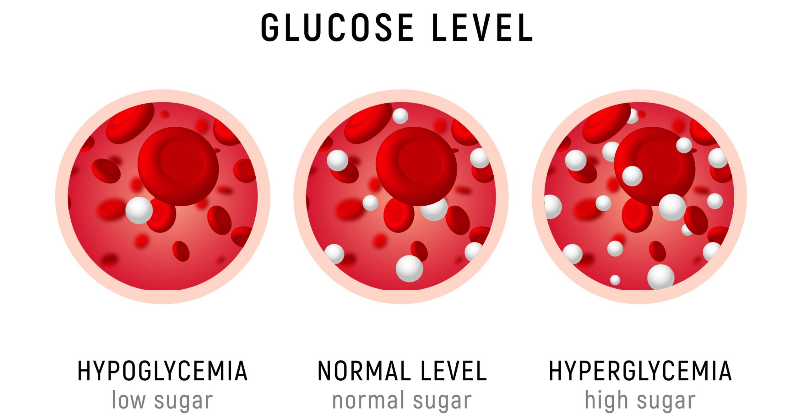
Hyperglycemia is marked by high glucose levels. In HHS, glucose levels can reach as high as 1500 mg/dL.
SIADH vs. Diabetes Insipidus
This video compares SIADH and diabetes insipidus, two disorders that affect fluid balance and serum sodium. The differential is relevant to HHS because clinicians may evaluate polyuria, hypernatremia, urine concentration, and mental status changes when deciding whether symptoms are due to HHS alone or another fluid and electrolyte disorder.
Diagnosis and Tests
Diagnosis of HHS is based on the patient’s history, physical examination, bedside glucose testing, and laboratory findings. Clinicians evaluate hyperglycemia, hyperosmolality, ketone production, acid-base status, kidney function, electrolyte loss, and the likely precipitating cause.
Typical diagnostic findings in HHS include:
- Plasma glucose usually >600 mg/dL
- Effective serum osmolality >300 mOsm/kg or total serum osmolality >320 mOsm/kg
- Absence of significant ketonemia; beta-hydroxybutyrate is usually <3.0 mmol/L
- Urine ketones may be negative, trace, or mildly positive but are usually <2+
- pH usually ≥7.3
- Bicarbonate usually ≥15 mmol/L
- Evidence of volume depletion, electrolyte shifts, and possible acute kidney injury
Common tests include a basic or comprehensive metabolic panel, serum osmolality, venous or arterial blood gas, beta-hydroxybutyrate, urinalysis, complete blood count, BUN and creatinine, magnesium, phosphate, ECG, chest imaging when indicated, and cultures if infection is suspected.
Because HHS can mimic or coexist with sepsis, stroke, renal failure, electrolyte disorders, and DKA, clinicians should reassess the diagnosis as new lab results and exam findings become available.
Management of Hyperosmolar Hyperglycemic Syndrome
Management of hyperosmolar hyperglycemic syndrome is an emergency, stepwise process focused on restoring circulating volume, correcting electrolytes, reducing glucose safely, treating the underlying trigger, and preventing complications from rapid osmolar shifts.
- Start with fluids. IV fluid resuscitation improves perfusion, supports kidney function, and begins lowering glucose by improving renal clearance.
- Check labs and electrolytes early. Monitor glucose, sodium, potassium, chloride, bicarbonate, BUN, creatinine, osmolality, ketones, and acid-base status.
- Replace potassium as needed before insulin. Insulin shifts potassium into cells and can worsen hypokalemia if potassium is already low.
- Begin insulin when it is safe. Insulin is usually started after initial fluid replacement and potassium assessment to avoid rapid osmolar shifts and dangerous potassium changes.
- Treat the trigger. Evaluate for infection, myocardial infarction, stroke, medication problems, missed diabetes therapy, or other precipitating illness.
- Monitor closely. Reassess hemodynamics, mental status, glucose, serum osmolality, kidney function, and electrolytes throughout treatment.
The Dangers of Hypovolemia
Hypovolemia is one of the most dangerous features of HHS. Severe hyperglycemia causes osmotic diuresis, which can lead to profound intravascular volume depletion.
Fluids come first because restoring circulating volume improves blood pressure, tissue perfusion, kidney function, and glucose clearance. Clinicians monitor hemodynamics, heart rate, blood pressure, capillary refill, urine output, serum sodium, serum osmolality, and mental status to judge whether perfusion is improving.
Dehydration worsens hyperosmolality because the blood becomes more concentrated as fluid losses continue. Careful fluid resuscitation and frequent reassessment help reduce the risk of neurologic deterioration from rapid or uncontrolled osmolar changes.
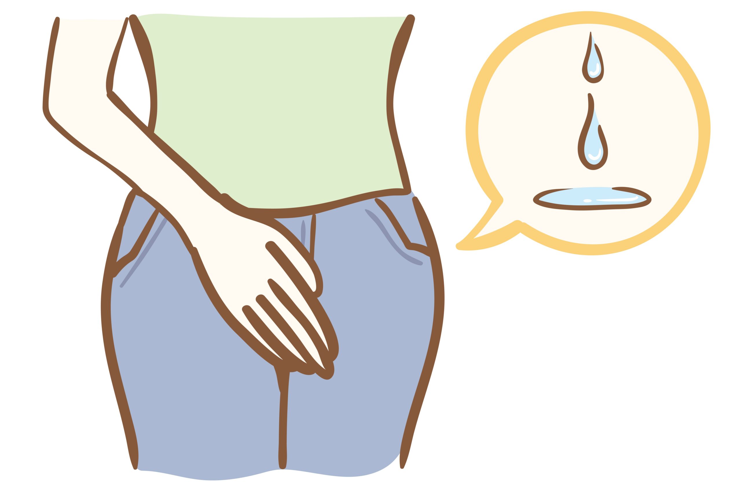
Frequent urination may lead to hypovolemia in patients presenting with HHS.
Minimal Ketones in Hyperosmolar Hyperglycemic Syndrome
HHS usually presents with minimal or absent ketones rather than the significant ketone production seen in DKA. However, absolute wording should be avoided because trace ketones or mild ketonemia may be present, and some patients have mixed DKA/HHS features.
Clinicians should also avoid assuming potassium is “not a concern” in HHS. Patients can have major total-body potassium depletion from osmotic diuresis, even if the initial serum potassium appears normal or high. Once insulin is started, potassium shifts into cells and hypokalemia can develop quickly.
The practical takeaway is to check ketones, pH, bicarbonate, anion gap, potassium, and osmolality so the team can distinguish HHS from DKA, recognize overlap, and treat safely.
Watch out for Hypokalemia
Providers must check potassium before starting insulin and continue monitoring it during treatment. HHS patients may be total-body potassium depleted because electrolytes are lost during osmotic diuresis, even when the first serum potassium result is normal or elevated.
The safety rule is simple: replace potassium as needed and do not start insulin if potassium is dangerously low until replacement has begun according to protocol. A patient on continuous IV insulin can develop falling potassium levels because insulin shifts potassium into cells, so understanding the regular insulin peak is important when monitoring electrolyte shifts and glucose reduction during treatment.
Potassium monitoring should be paired with frequent glucose checks, repeat electrolytes, kidney function monitoring, and reassessment of mental status and serum osmolality.
Summary
Hyperosmolar hyperglycemic syndrome is a life-threatening hyperglycemic state marked by severe hyperglycemia, hyperosmolality, and minimal or absent ketoacidosis. It is most often associated with type 2 diabetes, but mixed DKA/HHS presentations can occur and should be recognized early.
The most important triggers include infection, missed or inadequate diabetes therapy, newly recognized diabetes, myocardial infarction, stroke, surgery, trauma, and medication effects. Diagnosis depends on clinical assessment and laboratory testing, including glucose, electrolytes, osmolality, ketones, pH, bicarbonate, kidney function, and infection evaluation when indicated.
Treatment focuses on fluids first, careful potassium replacement, insulin when safe, trigger management, and close monitoring. Prognosis improves with early recognition, careful correction of fluid and electrolyte abnormalities, and prompt treatment of the underlying cause.
More Free Resources to Keep You at Your Best
Editorial Note
ACLS Certification Association (ACA) uses only high-quality medical resources and peer-reviewed studies to support the facts within our articles. Explore our editorial process to learn how our content reflects clinical accuracy and the latest best practices in medicine. As an ACA Authorized Training Center, all content is reviewed for medical accuracy by the ACA Medical Review Board.
More to Learn
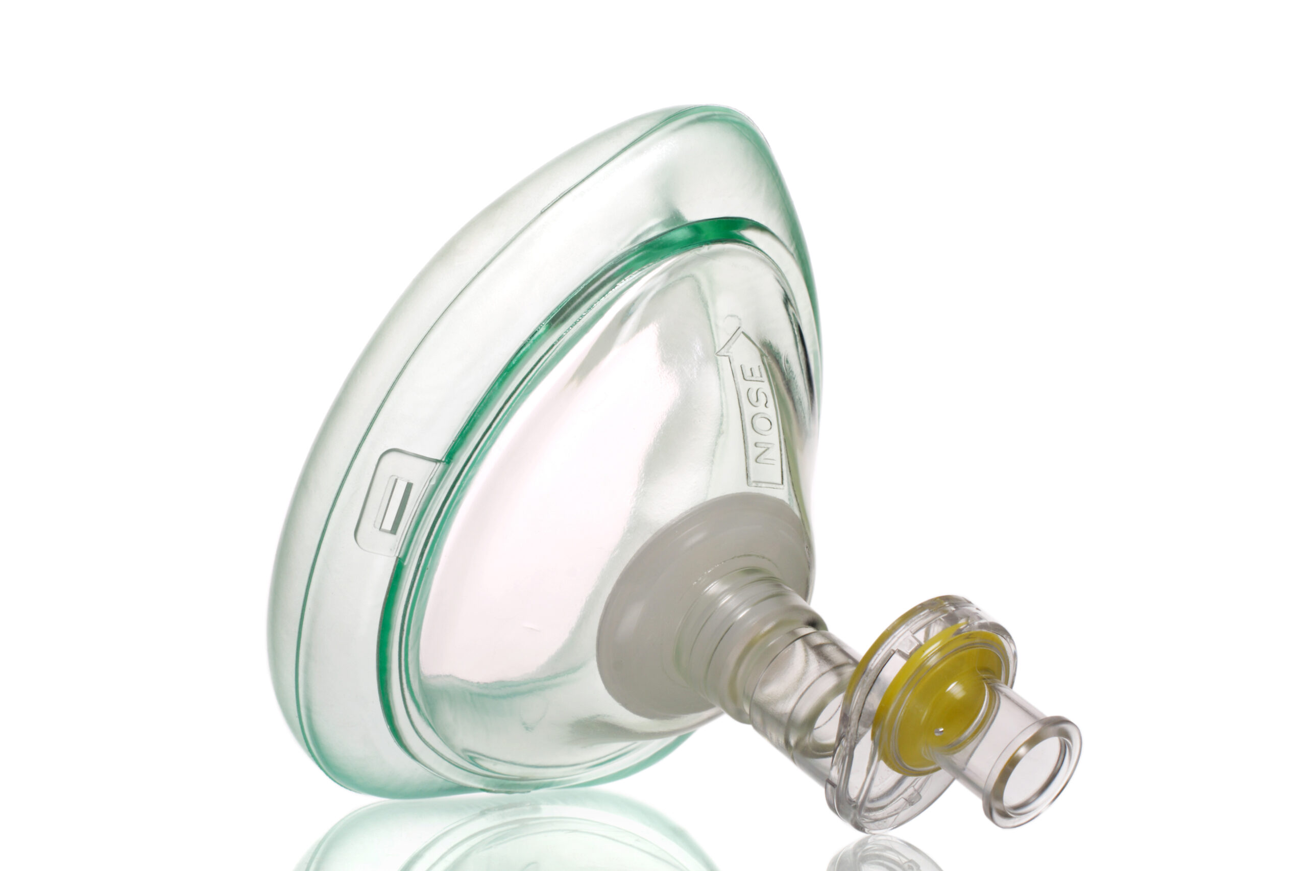
Effective ventilation is achieved by maintaining airway patency. Learn more about mouth-to-mouth ventilation and when available, a bag-mask device that is a more effective tool.

Understand Hypovolemia, a condition marked by a decrease in blood volume due to fluid loss or bleeding, through our informative article.