Criteria for Determining a Second-Degree Type 1 Heart Block
ACLS Certification Association videos have been peer-reviewed for medical accuracy by the ACA medical review board.
Article at a Glance
- Second-degree heart block Type I (Wenckebach) is characterized by:
- Regularity: R to R irregular with grouped beats
- Rate: Usually slower than normal
- P Wave: P waves are uniform but not always followed by QRS
- PR Interval: Becomes progressively longer until a P wave is blocked and the QRS is dropped
- QRS Complex: < 0.12 seconds
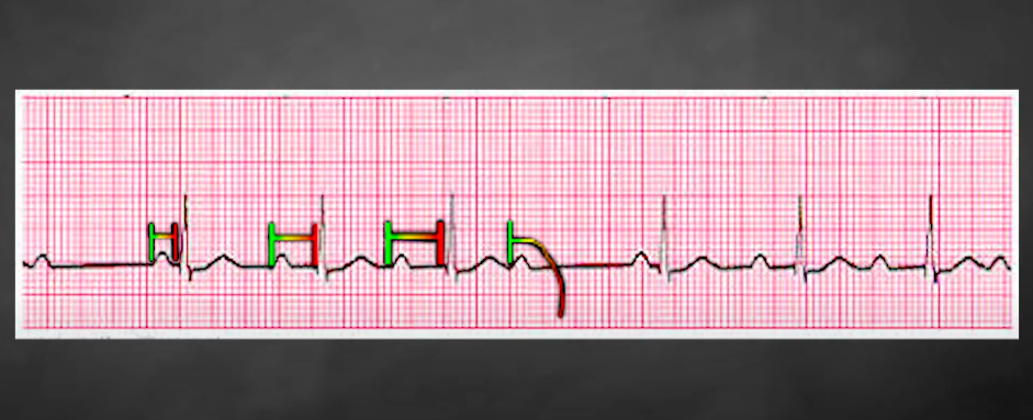
First, look at the relationship between the P wave and the QRS complex. In a second-degree Type I heart block, you’ll find an increasingly longer PR interval until the QRS complex drops off, unlike a first degree heart block where the PR interval is consistently prolonged but every QRS is conducted. The PR interval becomes increasingly longer until the QRS complex drops off. In the image above, the second PR interval is longer than the first and so on. The QRS complex drops off, and then it starts over. It’s the telltale sign for a second-degree type 1 heart block.Criteria to Determine a Second-Degree Type I Heart Block
Related Video – ECG and the Cardiac Cycle Basics
Summary
To diagnose a second-degree atrioventricular block, providers must interpret an ECG diagram, paying close attention to the P waves, PR interval, and QRS complex. During a second-degree type 1 heart block, the PR interval increases until the QRS complex drops off.
Related Video – What are Atrioventricular Blocks?
More Free Resources to Keep You at Your Best
Editorial Note
ACLS Certification Association (ACA) uses only high-quality medical resources and peer-reviewed studies to support the facts within our articles. Explore our editorial process to learn how our content reflects clinical accuracy and the latest best practices in medicine. As an ACA Authorized Training Center, all content is reviewed for medical accuracy by the ACA Medical Review Board.
More to Learn
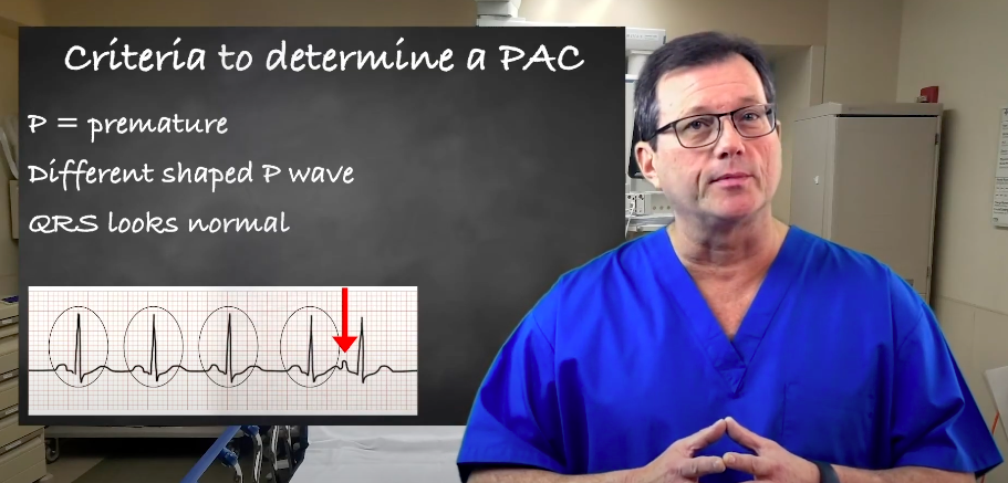
Explore premature atrial contractions (PACs) and the three determinants used to identify them. Our article will enhance your understanding of cardiac arrhythmias.
PEA is often caused by medical conditions that cause reversible cardiac arrest. Download the PDF and read our detailed article on Treating Reversible Causes, ROSV, and Terminating Resuscitation Efforts.