Care of Burn Patients
Burn patient care means stopping ongoing injury, assessing airway, breathing, circulation, protecting the wound, supporting perfusion, and deciding whether the patient needs burn-center care. This guide is for EMS providers, ED clinicians, and learners, and it follows the workflow from scene safety and pre-hospital care to emergency department assessment, resuscitation, transfer decisions, and early hospital treatment.
ACLS Certification Association videos have been peer-reviewed for medical accuracy by the ACA medical review board.
Article at a Glance
- Burn care starts with stopping the burning process while protecting rescuers and preventing additional injury.
- Airway, breathing, and circulation guide early priorities, especially when inhalation injury, burn shock, or associated trauma is possible.
- Pre-hospital care focuses on scene safety, ABCs, exposure control, IV access, pain control, rapid assessment, and transport.
- Hospital care adds reassessment, burn-depth and TBSA estimation, fluid resuscitation, transfer decisions, wound care, infection monitoring, pain control, and rehabilitation planning.
- Burn-center referral and fluid monitoring are key decision points, especially for a major burn, inhalation injury, high-risk burn locations, or inadequate urine output during resuscitation.
Burn Care Is Complex
There are three distinct phases of care for burn patients: the emergent phase, the hospital phase, and the long-term care phase.
The emergent, or pre-hospital, phase begins when healthcare providers arrive at the scene and continues through transport.
The hospital phase begins when the patient arrives at the emergency department and continues through the hospital or burn center stay.
The long-term care phase addresses recovery needs after discharge, especially for patients with severe burns.
Ongoing skin and wound care is individualized based on burn depth, location, size, infection risk, pain, mobility, and patient needs. Follow-up planning may include wound care instructions, scar management, contracture prevention, therapy, nutrition support, and return precautions.
How burns heal depends on depth and location. Superficial burns may heal with minimal intervention, while deeper burns skin, joints, hands, face, or circumferential areas may scar, tighten, limit function, or require reassessment if pain, swelling, drainage, fever, worsening redness, numbness, or reduced movement develops.
This guide focuses on the resuscitative phase, including the pre-hospital scene workflow and the early hospital phase.
The resuscitative phase includes the pre-hospital and hospital phases.
The care of burn patients is complex. These patients have many potential complications and injuries, such as burn shock, inhalation injury, associated trauma, infection, pain, and fluid loss. The care provided in the immediate first few hours after the burn injury is important to outcomes.
The resuscitative phase includes the pre-hospital and hospital phases.
The care of burn patients is complex. These patients have many potential complications and injuries, such as going into burn shock. The care provided in the immediate first few hours after the burn injury is paramount to successful outcomes.
Understanding Burn Treatment
This video gives a high-level overview of burn treatment priorities, including stopping the burning process, assessing the patient, and supporting early care. It fits here because the rest of the article breaks those priorities into scene and hospital steps.
Pre-hospital Phase (At the Scene)
The pre-hospital phase is an ordered checklist for the emergent, resuscitative phase of burn care. Providers first make the scene safe and stop ongoing injury, then move through airway breathing circulation, rapid assessment, history, and transport decisions.
In this phase, the provider must:
- Remove the patient from the burn source.
- Evaluate the patient’s airway and determine if there is a need to intubate.
- Address breathing and provide oxygen if needed.
- Address circulation, including removing clothing, starting an IV, and administering intravenous fluids.
- Perform a quick head-to-toe assessment.
- Obtain patient history.
These steps are discussed further below.
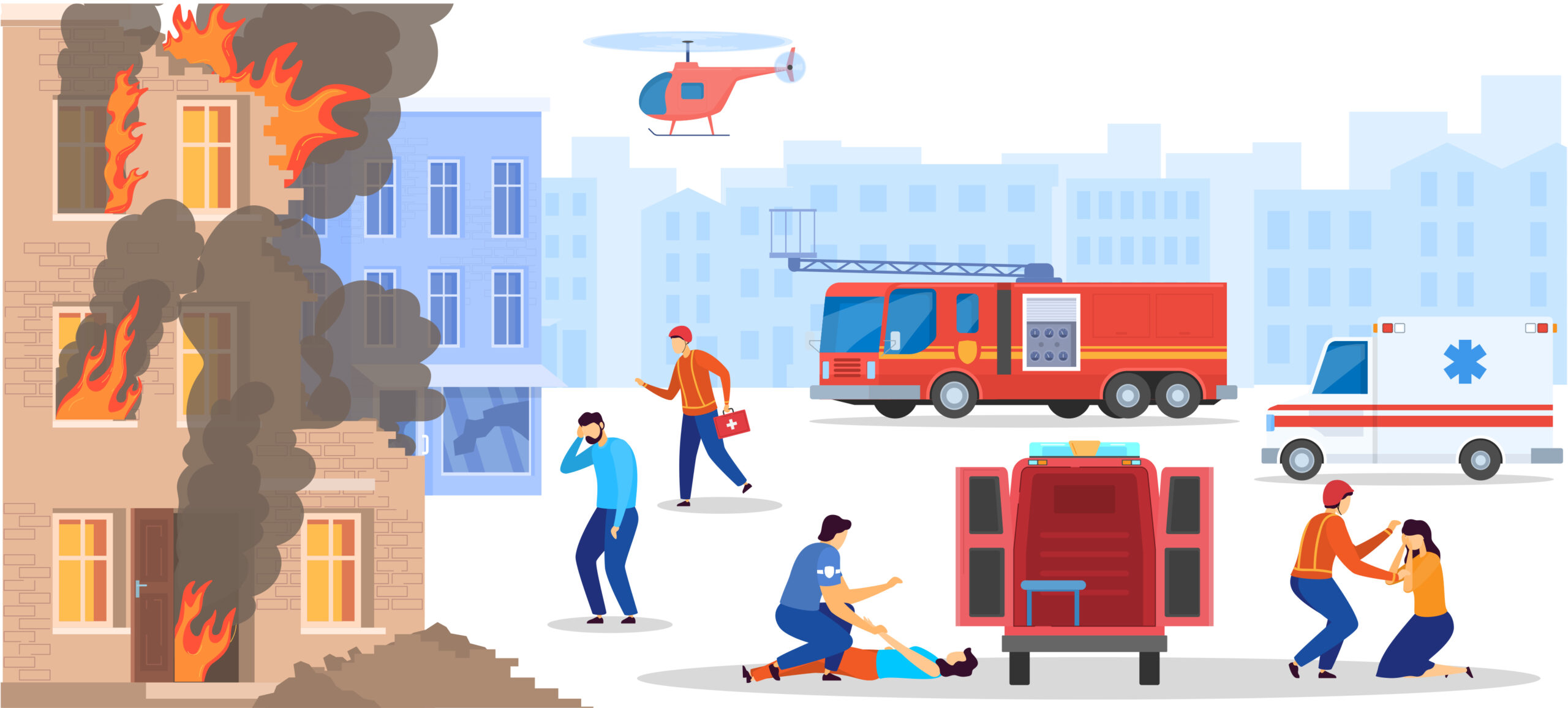
For burn patients, the first priority is to stop the burning process, such as removing a patient from a building on fire.
Stop the Burning Process
The first priority of the healthcare provider when arriving at the scene is to stop the burning process. Because the burn is causing the injury, it needs to be stopped as quickly as possible. However, the way to stop the process depends on the type of burn: thermal, chemical, electrical, or scalding.
Thermal Burns
For a thermal burn, the flames must first be extinguished, which may require smothering them with a blanket or water. Ice should never be used to stop a burn, as it causes significant tissue damage.
Chemical Burns
The first step to stopping a chemical burn is to ensure no chemicals or powders are in contact with the patient. That may require removing the patient’s clothing immediately to eliminate the source of contact. Once the source has been removed, the patient should be doused with water.
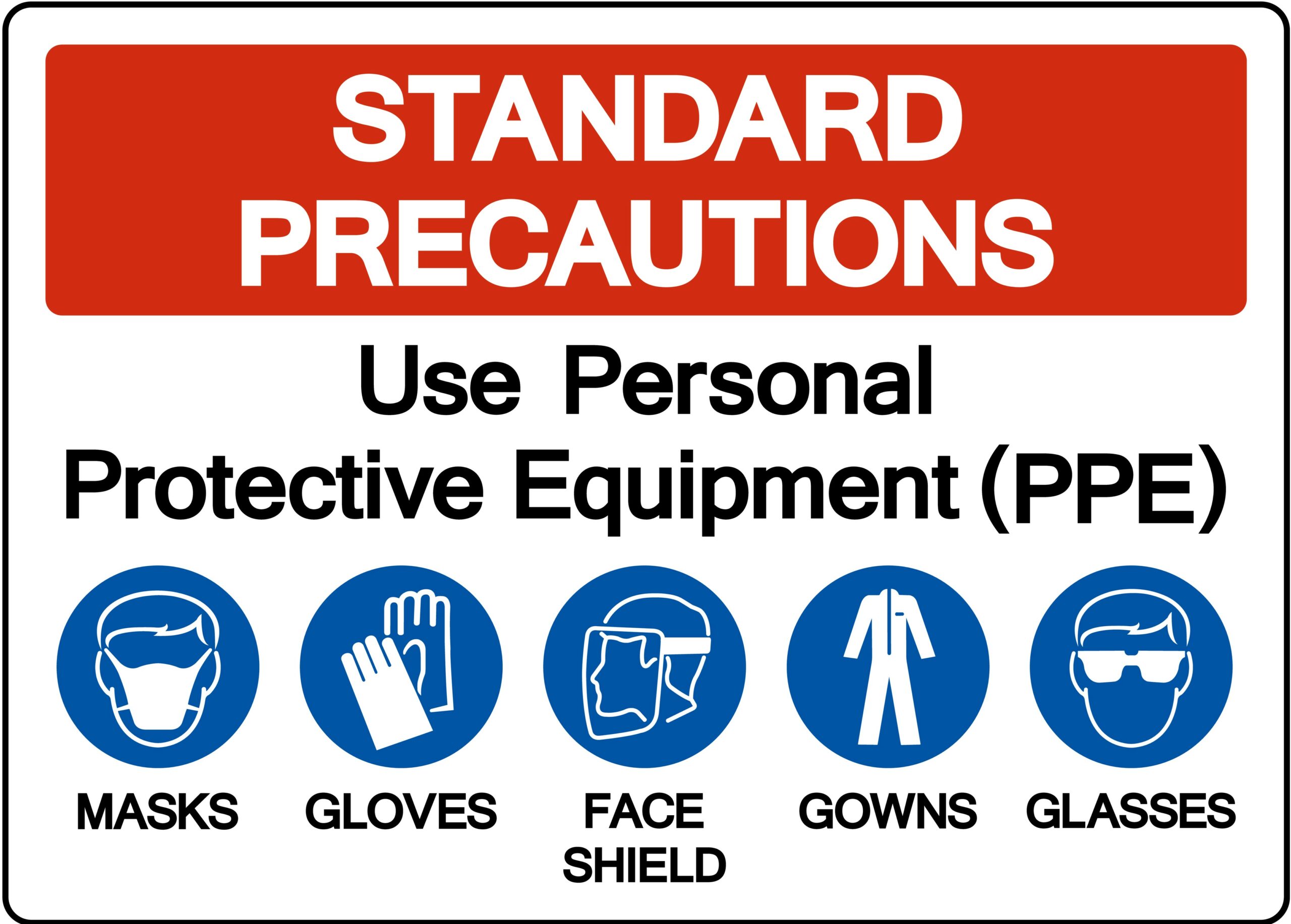
When dealing with chemicals or a chemical burn, the provider must wear personal protective equipment (PPE) to avoid coming in contact with the chemical.
Electrical Burns
For an electrical burn, the patient needs to be removed from the electrical source. Any providers on the scene must take precautions to avoid coming in contact with the electrical source themselves.
Scalding Burns
If the patient is scalded by something like hot tar or asphalt, they first need to be removed from the source of the scalding. If clothing is stuck to them, the provider should not attempt to remove it, as skin and tissue could be removed as well.
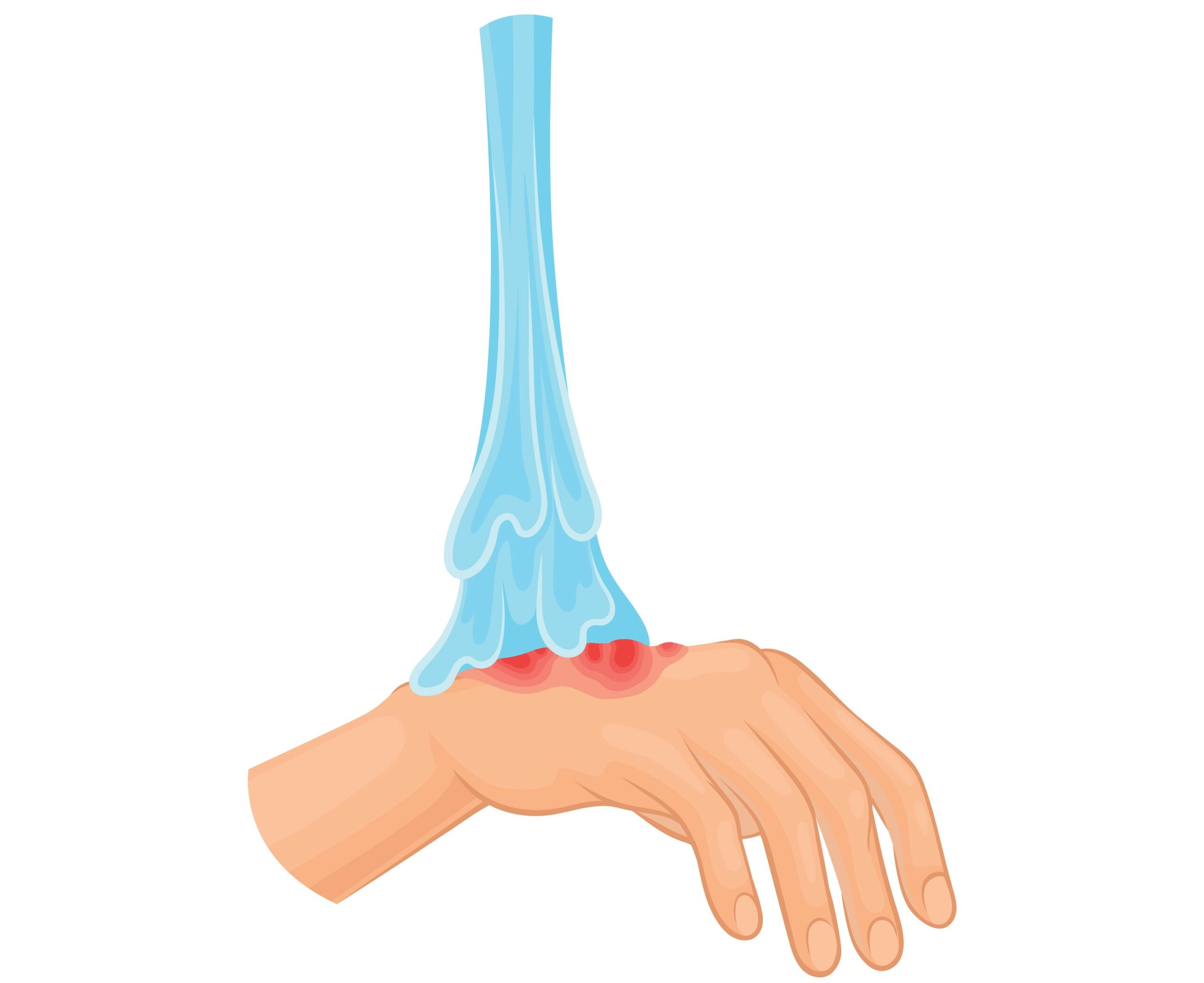
For a chemical burn, remove the clothing and douse the burn with water.
After ongoing injury is stopped, transition immediately to ABCs, exposure control, pain management, and transport planning. Cooling and irrigation can be helpful in selected burns, but avoid prolonged exposure, ice, or excessive cooling that could worsen hypothermia risk, especially while waiting for transport.
Assess the Airway
Once the burning process is stopped, it is essential to assess the airway.
Patients with significant facial burns, soot in the mouth or sputum, hoarseness, stridor, respiratory distress, or suspected inhalation injury have a higher risk of airway edema. Airway swelling can progress over time, so early airway-control decisions are often made before edema worsens and intubation becomes more difficult.
Once the patient has been intubated or an oral airway has been put into place, it is essential to address breathing with oxygen.
Address Breathing with Oxygen
For a patient with smoke inhalation, inhalation injury, or evidence of airway edema, 100% humidified oxygen should be administered through the endotracheal (ET) tube or a 100% non-rebreather mask at the scene.
Address Circulation
After assessing the airway and administering oxygen on the scene, circulation must be addressed. Any clothing or jewelry that is restricting the patient from perfusing or breathing needs to be removed.
Large-bore IVs, either 18 or 16 gauge, should be started to administer fluids. The fluid of choice is lactated Ringer.
Perform a Quick Head-to-Toe Assessment
A rapid head-to-toe assessment should be completed to assess for any other life-threatening injuries before arriving at the hospital.
A rapid head-to-toe assessment assesses the patient’s neurological status and any other life-threatening injuries and circumferential burns.
Part of the head-to-toe assessment is to assess the patient’s spine to determine if cervical spine precautions are needed for transport. If the patient fell from a height or has a suspected spinal injury, they will need to be placed on a backboard.
If the patient is suspected of having a spinal injury, narcotic pain medication may be administered through the IV to help manage pain during transport.
Understanding Chest Tubes
This video is relevant when burn patients also have associated trauma, blast injury, rib fractures, pneumothorax, hemothorax, or respiratory complications that require chest tube placement. It should be understood as related trauma-support content rather than routine care for every burn patient.
Obtain a Brief History from the Patient
Because the patient will likely be awake and alert at the scene, the provider needs to obtain a brief patient history. Once at the hospital, the patient may be sedated or rushed to surgery, making it difficult for them to communicate with the medical team.
Questions the provider should ask include:
- What happened?
- What type of burn is it?
- What led to the burn?
- What chemicals were involved?
- What is your medical history?
- Are you allergic to any medications or anything else?
- What medications do you take?
- What diseases do you have that could complicate anesthesia?
Having information on the patient’s history is essential for the continuum of care at the hospital.
Once these steps have been taken, the patient is ready for transport to the hospital.
Read: Three Common Types of Pneumonia
Hospital Phase
Upon arrival at the hospital, the patient’s ABCs (airway, breathing, and circulation) will continue to be monitored and assessed. Because burn patients may require repeated hands-on care, airway support, invasive devices, and wound-related precautions, clinicians should also follow appropriate infection control and isolation practices according to facility policy.
Airway
Patients who were not intubated at the scene will be further assessed for new stridor, wheezing, and evidence of airway edema. If any of these are present, the patient will need to be intubated.
For patients who were intubated at the scene, an X-ray should be obtained to confirm the placement of the ET tube. The provider must also confirm that the ET tube is well secured, as it could be difficult to reintubate due to edema or inhalation injury should the patient be accidentally extubated.
Care must be taken when securing the ET tube to ensure that it is not secured to any area with a burn injury (such as around the ears) to avoid causing more tissue injury.
Unlike at the scene, the provider at the hospital has access to more equipment to better assess the patient. For example, a fiber optic bronchoscope may be used to further check the patient’s airway and visualize airway injury.
Once the airway is secured, the provider must assess the patient’s breathing.
Breathing
The provider assesses the patient’s breathing by listening to breath sounds, assessing the quality of the patient’s respirations, and examining the sputum (e.g., is it tarry or bloody? Is the patient coughing up soot? Is there soot around the patient’s mouth?).
The provider will also assess for chest wall excursion, examining for evidence of flail chest.
If carbon monoxide exposure is suspected, clinicians may assess carboxyhemoglobin levels and recognize that standard pulse oximetry can be falsely reassuring. High-flow oxygen or 100% FiO2 is commonly used while the team evaluates severity and local treatment options.
If cyanide exposure is suspected, clinicians may assess lactate, acid-base status, and clinical signs of severe toxicity; cyanide levels are often not rapidly available in the acute setting. Antidotal therapy, such as hydroxocobalamin, may be considered when suspicion is high according to local protocol.
In addition to administering oxygen to assist with breathing, medications, such as albuterol, can be administered to help open the patient’s airways.
Once the patient’s breathing is assessed, it is important to next address the patient’s circulation.
Circulation
While assessing circulation, the provider needs to estimate the total body surface area that has been burned using the Rule of Nines.
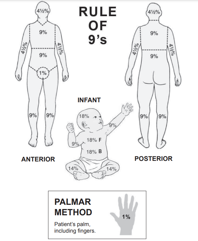
The Rule of Nines estimates the total body surface area affected by burns.
The Rule of Nines is a guide providers use to estimate the total burned body surface area. Patients with a significant percentage of burned surface area will need to be transferred to a burn center for continued care.
Burn-center consultation or transfer should follow local policy and burn-center guidance. The American Burn Association recommends immediate consultation with consideration for transfer for categories such as:
- Full-thickness burns
- Partial-thickness burns involving 10% or more of total body surface area (TBSA)
- Any deep partial-thickness or full-thickness burns involving the face, hands, feet, genitalia, perineum, or over major joints
- Suspected inhalation injury
- Chemical injuries
- High-voltage electrical injuries or lightning injury
- Burns with major trauma, significant comorbidities, uncontrolled pain, or special pediatric needs
These criteria are intended to support referral decisions, not replace clinical judgment. See the American Burn Association burn patient referral guidelines for the full referral framework.
Using the Parkland Formula to Estimate IV Fluid Replacement
Burn patients require significant amounts of fluid because of capillary permeability and extensive fluid loss from the intravascular space. If the fluid is not adequately replaced, they will experience burn shock. The provider uses the Parkland formula to estimate how much fluid the patient needs.
The Parkland formula is:
Fluid requirement=Body surface area burned % × weight kg× 4 mL
Note: the 4 mL variable may vary between healthcare organizations.
For example, if the total burned surface area is 25% and the patient weighs 100 kilograms, the formula is: 25 × 100 × 4 = 10,000. (Note: 25 is used for the percentage, not 0.25.)
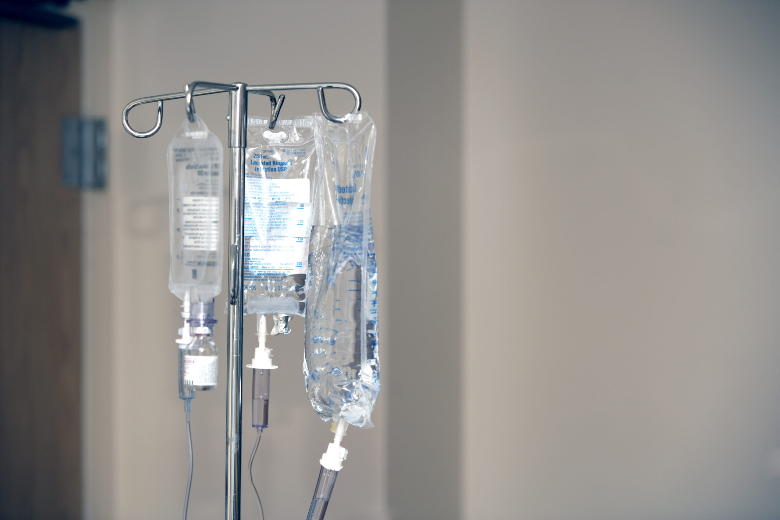
IV fluid replacement is crucial for burn patients.
As illustrated in the example above, the Parkland formula will return a large number for fluid replacement (e.g., 10,000 mL). Burn patients require a lot of fluid.
The Parkland formula estimates the total crystalloid fluid requirement for the first 24 hours after the burn. Using the example above, the patient requires 10,000 mL of fluid replacement in the first 24 hours.
Standard Parkland timing gives half of the calculated volume in the first 8 hours from the time of the burn, not from hospital arrival, and the remaining half over the next 16 hours. In this example, 5,000 mL would be targeted for the first 8 hours from burn time, and 5,000 mL over the following 16 hours.
Formula-based fluid estimates are starting points. Fluid rates must be titrated to patient response, urine output, hemodynamics, burn size reassessment, inhalation injury, associated trauma, age, and local protocol.
Reference: NCBI Bookshelf: Burn Fluid Resuscitation.
Checking if Fluid Replacement is Adequate
Once fluid replacement is initiated, clinicians monitor whether resuscitation is adequate. A Foley catheter allows urine output to be measured and used with vital signs, mental status, perfusion, lactate or base deficit when available, and overall clinical response.
For many adult burn resuscitation protocols, a general urine output reference is about 30–50 mL/hour or 0.5–1 mL/kg/hour. This target is not universal; children, smaller adults, electrical burns, rhabdomyolysis, renal injury, and local burn-center protocols may require different goals.
Burn Care Treatment Options
Hospital burn care is individualized by burn depth, TBSA, location, airway risk, comorbidities, pain, infection risk, mobility, and whether the patient needs a burn center.
Wound Coverage and Dressings
Cover the wound with clean, dry dressings or burn-specific dressings according to facility policy. Wound coverage helps reduce contamination, heat loss, pain, and further tissue exposure.
Topical Agents
Topical antimicrobial or moisture-balancing agents may be used when indicated. Selection depends on burn depth, location, infection risk, allergy history, and burn-center or wound-care guidance.
Debridement
Debridement may be needed to remove devitalized tissue, reduce contamination, or prepare the wound bed for healing or procedures. Timing and technique depend on burn depth, stability, and specialist input.
Infection Monitoring and Antibiotics When Indicated
Clinicians monitor for fever, worsening pain, spreading redness, swelling, purulent drainage, odor, cellulitis, sepsis, or graft failure. Antibiotics are not used automatically for every burn, but they may be started when infection is suspected or confirmed.
Pain and Anxiety Control
Burn care can be extremely painful, especially during dressing changes, transport, procedures, and therapy. Pain and anxiety control should be planned, reassessed, and adjusted to the patient’s condition and airway risk.
Tetanus Prophylaxis
Tetanus immunization status should be reviewed, and prophylaxis should be provided according to wound type, immunization history, and facility protocol.
Therapy and Rehabilitation
Early physical and occupational therapy may help preserve range of motion, reduce contracture risk, support positioning and splinting, and improve long-term function.
Procedures, Skin Graft, and Pressure Relief
Deep burns may require excision, skin graft procedures, or other operative care. Circumferential burns of the chest, limbs, or digits may require pressure-relieving procedures, such as escharotomy, when perfusion or ventilation is compromised.
Summary
Burn patient care begins with scene safety, stopping the burning process, preventing hypothermia, and moving quickly through airway, breathing, circulation, rapid assessment, history, and transport. In the hospital, clinicians reassess ABCs, evaluate inhalation injury, estimate burn size and depth, begin or adjust fluid resuscitation, monitor urine output, manage pain, protect the wound, and decide whether burn-center consultation or transfer is needed.
Care does not end after initial stabilization. Burn patients may need wound care, dressings, topical therapy, debridement, infection monitoring, tetanus prophylaxis, rehabilitation, scar and contracture prevention, grafting, or pressure-relieving procedures depending on injury severity. The next step is to follow local burn protocols and involve a burn center early when referral criteria or clinical concern are present.
More Free Resources to Keep You at Your Best
Editorial Note
ACLS Certification Association (ACA) uses only high-quality medical resources and peer-reviewed studies to support the facts within our articles. Explore our editorial process to learn how our content reflects clinical accuracy and the latest best practices in medicine. As an ACA Authorized Training Center, all content is reviewed for medical accuracy by the ACA Medical Review Board.
More to Learn
Understand the differences between CPR and defibrillation by reading our article. Learn about their critical roles in cardiopulmonary resuscitation and life-saving interventions.
This article and video explain Isolation and Infection Precautions. Understand the personal protective equipment for standard, contact...