Three Common Types of Pneumonia
ACLS Certification Association videos have been peer-reviewed for medical accuracy by the ACA medical review board.
Article at a Glance
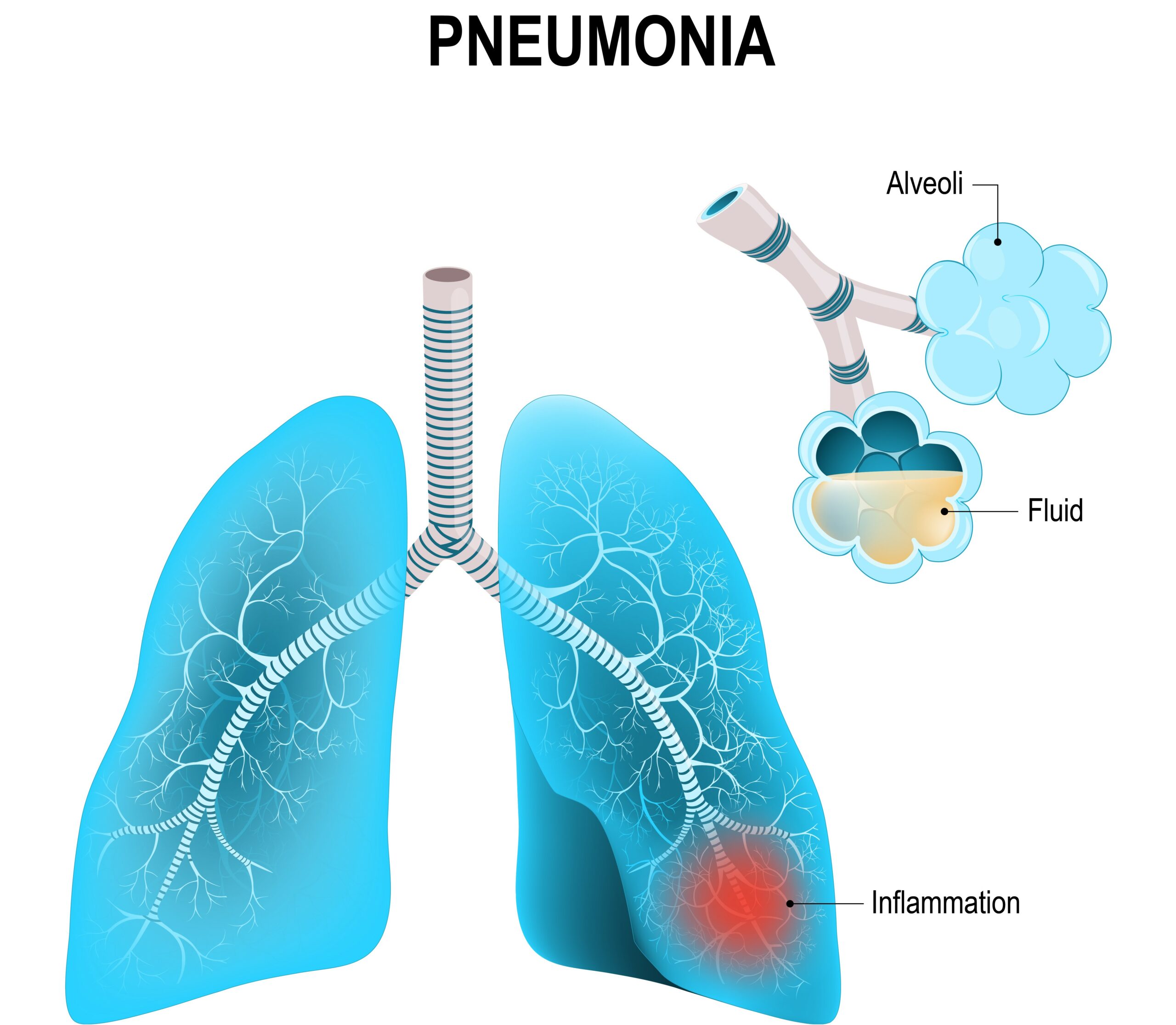
- Pneumonia is an infection causing inflammation in one or both lungs with fluid and pus build-up in the alveoli.
- Three common types of pneumonia are lobar, bronchopneumonia, and interstitial pneumonia.
- Clinicians will learn about the risk factors and unique signs and symptoms for different types of pneumonia.

Pneumonia causes pus or fluid build-up in the alveoli.
The risk factors for pneumonia are:1 A decreased level of consciousness may cause aspiration pneumonia because patients with a decreased LOC are at risk for aspiration. Trouble coughing up and clearing secretions is a prominent contributing factor to pneumonia. Another risk factor is intubation with mechanical ventilation. When a patient is on a ventilator, they are not able to protect their airway or cough up secretions. They rely on the nurse to suction the airway clean. Many hospitals have implemented a “ventilator bundle” — a protocol nurses and respiratory therapists follow to help prevent patients from contracting ventilator-associated pneumonia. The protocol includes keeping the head of the bed raised, performing oral care every two hours, suctioning as needed, gastrointestinal (GI) prophylaxis, deep vein thrombosis (DVT) prophylaxis, and turning every two hours. The elderly are also at risk for pneumonia. They have decreased immune responses due to age and are more at risk for developing pneumonia as a secondary infection. If a 75-year-old patient with diabetes and heart disease contracts a sinus infection, they’re at a much higher risk for the infection developing into pneumonia compared to a healthy 20-year-old2. Smoking decreases the action of the cilia within your respiratory tract. Cilia are little hair-like tendrils that help move mucus up and out, and decreased ciliary action increases the risk for pneumonia. Finally, patients with chronic diseases such as diabetes, congestive heart failure, cancer, and autoimmune diseases are at a higher risk of developing pneumonia as well as patients receiving chemotherapy, high-dose steroids, and immunosuppressants.Risk Factors of Pneumonia
Related Video – How to Manage Respiratory Arrest on an Adult
There are three main types of pneumonia. They have slightly different causes and present differently. They are: Read: Care of Burn PatientsThree Main Types of Pneumonia
Lobar
Bronchopneumonia
Interstitial
Caused By:
S. pneumoniae
Aspiration (multiple bacteria)
Fungus, opportunistic infection (PCP)
Distinguishing Signs & Symptoms:
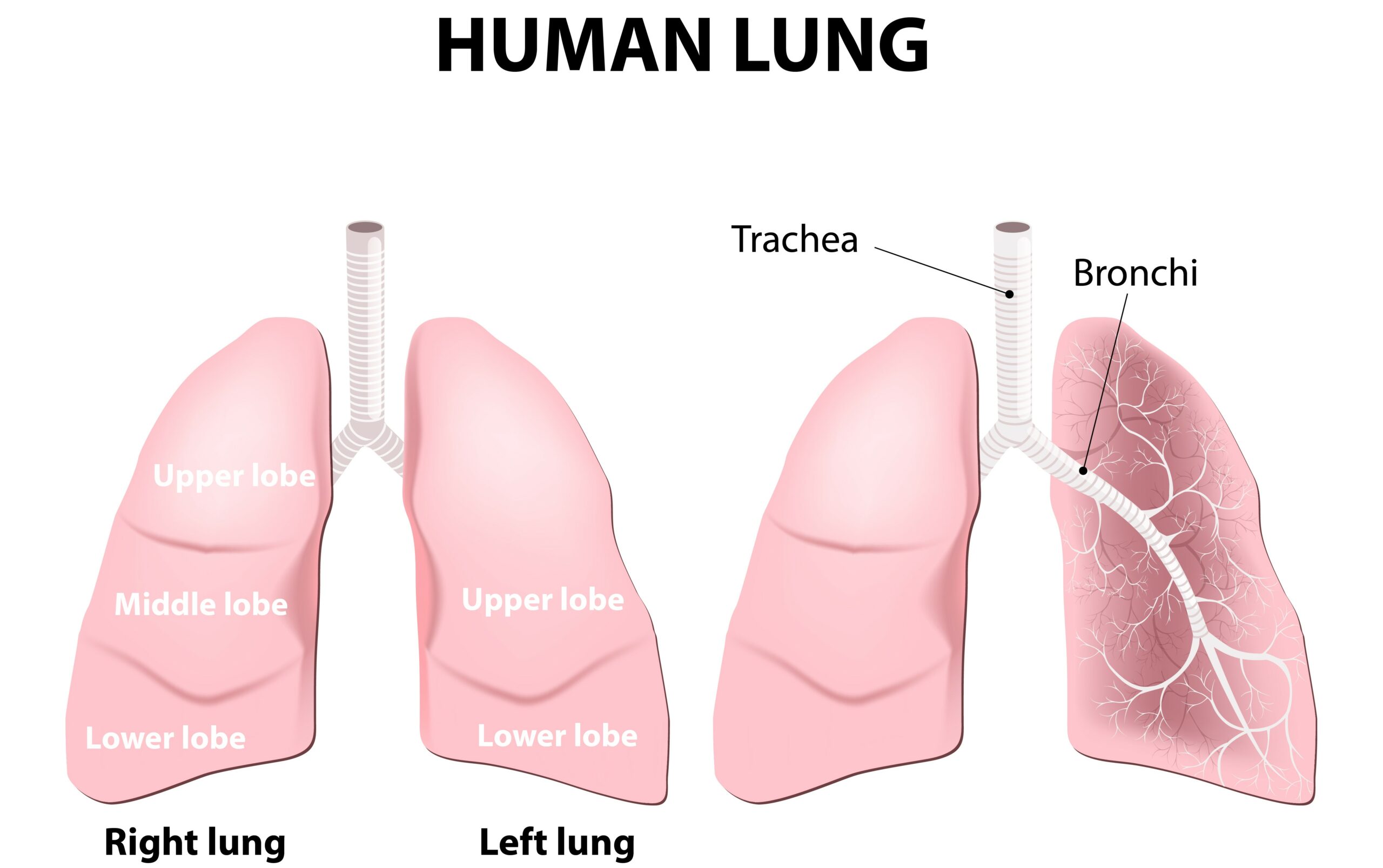
Lobar pneumonia is the most common type. It is also called pneumococcal pneumonia because the most common bacteria responsible is called streptococcus pneumoniae (also known as S. pneumoniae or strep pneumoniae). Clinicians administer antibiotics to patients with lobar because it’s a bacterial infection. It’s generally the easiest to treat and least deadly type of pneumonia. It’s paramount that clinicians understand how each type of pneumonia presents. Each has unique disease processes, which make it easier to differentiate. Patients with pneumonia will cough, but clinicians must diagnose the type of cough, color of sputum, and types of breath sounds. Rusty color sputum is unique to lobar pneumonia. Bacteria flood the alveoli, causing an inflammatory response that damages the alveolar capillaries. White blood cells and red blood cells leak into the airway, creating the rusty color. A physician will hear crackles due to alveolar collapse from the fluid build-up and bronchial breath sounds instead of Broncho vesicular sounds over the affected lung field. For example, right middle lobe pneumonia will eventually cause fluid consolidation in the right middle lobe. In lobar pneumonia, consolidation may be limited to just one lobe or multiple. As the fluid consolidates and settles into one lobe, it acts as a sound magnifier. Clinicians will hear magnified breath sounds around the affected lobe. Abnormal, deep bronchial breath sounds may be present in the affected lung region as opposed to normal Broncho vesicular sounds. The consolidation causes sounds to bounce off the fluid, making the lung sounds more bronchial on auscultation. An X-ray will also display a consolidation.Lobar
Signs and Symptoms

Related Video – Respiratory Drugs – Part 1
Aspiration is the most common cause of bronchopneumonia. For example, a patient with a stroke or decreased LOC may aspirate. Aspiration leaves patients open to a plethora of bacteria, and when a patient aspirates vomit or food, multiple bacteria may grow. It is more difficult to treat bronchopneumonia because multiple bacteria are involved. The bacteria cause a yellowish-green sputum unique to bronchopneumonia.Bronchopneumonia
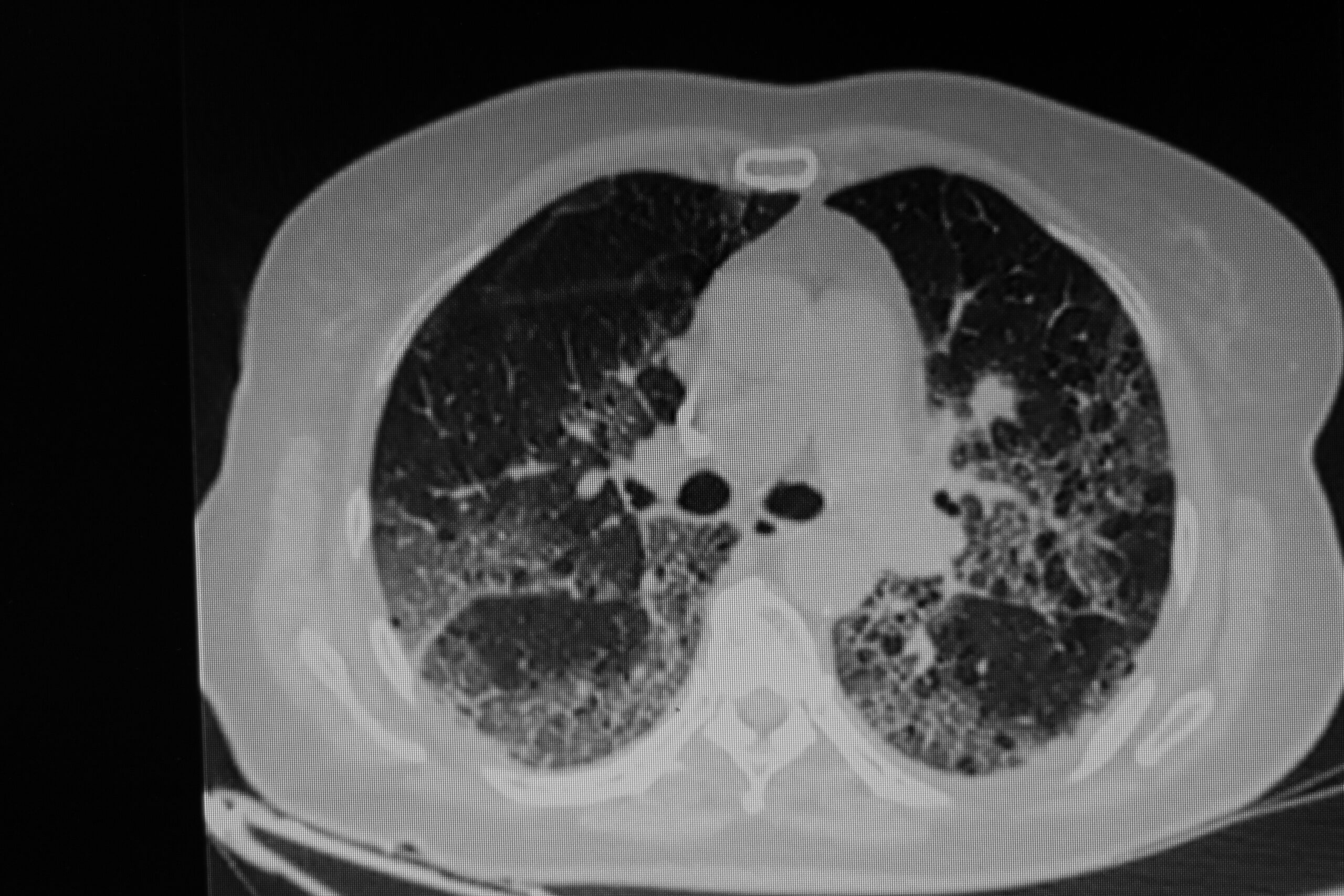
Interstitial pneumonia is a group of lung diseases that cause fluid build-up in the interstitial space. Pneumocystis pneumonia (PCP) is a common cause of interstitial pneumonia.. PCP has a high mortality rate and is difficult to treat. It mostly affects the immunocompromised, such as patients with HIV/AIDS or on high-dose immunosuppressants. The fungus pneumocystis jirovecii (formerly called pneumocystis carinii) causes PCP. PCP is a serious complication of AIDS and can be deadly. The AIDS HIV virus itself kills very few patients; most deaths in AIDS patients are caused by an infection the AIDS patient contracts… Opportunistic pathogens are common in patients with weakened immune systems. A non-productive, dry cough with little to no mucus is one of PCP’s unique symptoms. The fungus invades the alveoli, causing an inflammatory response that results in fluid leakage. The alveoli fill up with fluid, which causes alveolar necrosis and kills off the alveoli. The patient’s non-productive cough is due to their irritated respiratory tract, which tries to expel the foreign substance. Interstitial pneumonia expresses as a ground glass appearance on a chest X-ray, typically appearing hazy and diffuse. It sometimes presents as a honeycomb because the pneumonia isn’t consolidated to one lobe or lung. A chest X-ray highlighting ground-glass opacities. The three basic types of pneumonia are lobar, bronchopneumonia, and interstitial pneumonia. They each present unique signs and symptoms, and clinicians should pay attention to the patient’s cough type and sputum color. Lobar is the least fatal and most easily treatable, and interstitial is the most difficult.Interstitial Pneumonia
Signs and Symptoms

More Free Resources to Keep You at Your Best
Editorial Sources
ACLS Certification Association (ACA) uses only high-quality medical resources and peer-reviewed studies to support the facts within our articles. Explore our editorial process to learn how our content reflects clinical accuracy and the latest best practices in medicine. As an ACA Authorized Training Center, all content is reviewed for medical accuracy by the ACA Medical Review Board.
1. Torres A, Peetermans WE, Viegi G, Blasi F. Risk factors for community-acquired pneumonia in adults in Europe: a literature review. Thorax. 2013;68(11):1057-1065. doi:10.1136/thoraxjnl-2013-204282
2. Henig O, Kaye KS. Bacterial Pneumonia in Older Adults. Infect Dis Clin North Am. 2017;31(4):689-713. doi:10.1016/j.idc.2017.07.015
