Interpreting a 12-Lead Electrocardiogram
ACLS Certification Association videos have been peer-reviewed for medical accuracy by the ACA medical review board.
Article at a Glance
- A 12-lead electrocardiogram records the heart’s electrical activity from 12 different areas.
- The right coronary artery and left coronary artery are the key places affected during myocardial infarctions.
- Clinicians will learn how to look for a STEMI on a 12-lead electrocardiogram.
The 12-lead electrocardiogram (ECG) is an essential diagnostic tool. Clinicians must evaluate a 12-lead ECG to look for myocardial infarction (STEMI) in each of the 12 leads. Electrocardiogram Lead Equipment The diagram below illustrates approximate lead placement. Each lead provides a different picture of the heart, allowing clinicians to observe heart activity from 12 angles. The 12 leads are placed at specific locations on the body. Clinicians check if the patient has a STEMI or elevated ST segment in each of the 12 leads. A STEMI is an ST elevated myocardial infarction. The ST segment is elevated. Read: An Easy Way to Interpret Arterial Blood GasesIntroduction to 12-Lead Electrocardiograms


The three inferior leads are II, III, and AVF. They’re located at the bottom of the heart. It’s where clinicians first look for a STEMI. If a STEMI is present in one or more of the inferior leads, it usually indicates a blockage somewhere in the right main coronary artery. The coronary arteries supply blood to the heart. Blockage in the coronary arteries can cause myocardial infarction. When a patient has a STEMI in II, III, or AVF, clinicians must rule out the possibility of a right ventricular infarction before administering vasodilators like morphine and nitroglycerin. If the patient has a right ventricular infarction, their preload is significantly reduced and administering vasodilators may lead to complications.Inferior Leads

Related Video – One Quick Question: How Can I Remember Contiguous 12-Lead ECG?
The next leads are I and AVL. They look at the high lateral wall. The “L” in AVL stands for “left.” If there is an ST elevation in lead I or AVL, the patient has a high lateral STEMI. In this case, there’s a blockage somewhere in the left main artery.Lateral Leads
Clinicians last look for a STEMI via the chest leads, also called precordial leads. All of the chest leads are placed across the heart. There are six of these “V” leads: V1, V2, V3, V4, V5, and V6. A STEMI typically affects a combination of areas in the heart. For example, a patient with a STEMI in V1, V2, and V3 has an anteroseptal myocardial infarction. The patient has STEMI in both the anterior section and the septal section. If all the V, AVL, and I leads are involved, the patient has a massive anterior myocardial infarction. A 12-lead electrocardiogram informs physicians of the heart’s electrical activity from 12 different angles.Chest Leads (Precordial Leads)

Related Video – Chest Pain EKG Interpretation for ACS
A 12-lead ECG measures heart activity from 12 different angles. Clinicians begin with the inferior leads, then the lateral leads, and finally the chest leads, making sure to rule out STEMIs as they progress.Summary
More Free Resources to Keep You at Your Best
Editorial Note
ACLS Certification Association (ACA) uses only high-quality medical resources and peer-reviewed studies to support the facts within our articles. Explore our editorial process to learn how our content reflects clinical accuracy and the latest best practices in medicine. As an ACA Authorized Training Center, all content is reviewed for medical accuracy by the ACA Medical Review Board.
More to Learn
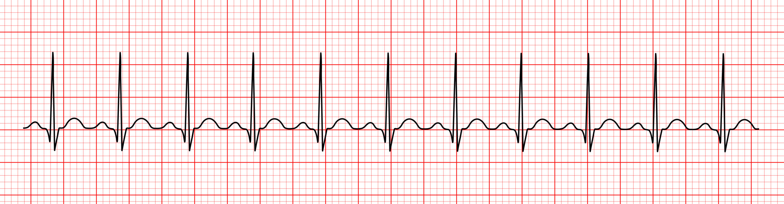
When interpreting the ECG strip, consider the following: (1) regularity, (2) rate, (3) the P-wave, (4) PR-interval, and (5) QRS-complex. Learn more in the detailed article.
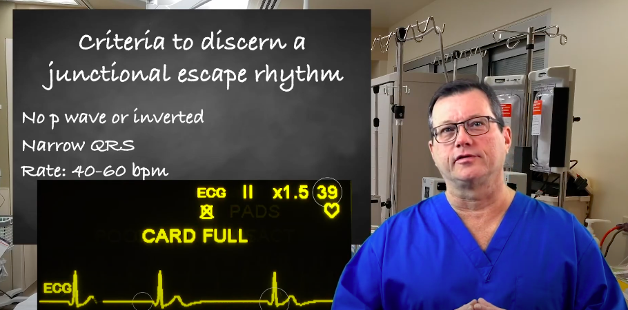
Learn about junctional escape rhythm criteria for ACLS certification with our article. Understand the guidelines and requirements seamlessly.