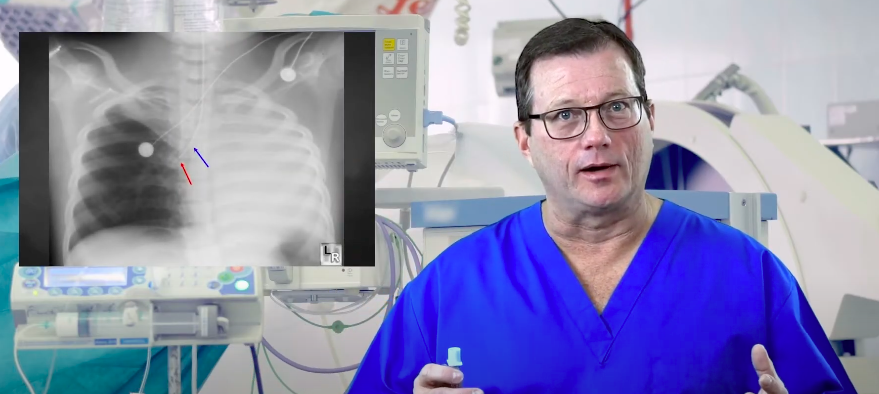
Hemodynamics: Part 2
ACLS Certification Association videos have been peer-reviewed for medical accuracy by the ACA medical review board.
Article at a Glance
- Some common hemodynamic measurements are central venous pressure, cardiac output, cardiac index, systemic vascular resistance, and pulmonary capillary wedge pressure.
- A catheter measures the pressure in different sections of the heart.
- Clinicians will learn about different factors affecting hemodynamic measurements.
In order to understand the intricacies of hemodynamics and to understand how conditions affect different hemodynamic measurements, clinicians must know cardiac anatomy and how blood flows through the heart. A summary of common hemodynamic measurements and their normal values. There are five hemodynamic measurements: The clinician should measure the pressure in different areas of the heart to understand how the structures are functioning.Understanding Hemodynamics


Related Video – Hemodynamics – Part 1
Central venous pressure (CVP) is a direct measurement of the pressure in the right atrium. Normally, CVP is between 2 to 6 mm Hg. CVP is sometimes referred to as right atrial pressure or preload.. The terms are interchangeable because preload is the result of venous return coming back to the right side of the heart, and CVP measures the venous return. Clinicians measure CVP using either the distal port of a central line (also known as a central venous catheter) or a triple lumen pulmonary artery catheter. CVP is the only measurement a clinician can make without placing a pulmonary artery catheter. Hypovolemia reduces CVP. A patient’s CVP decreases when the volume coming back to the right side of the heart is decreased, causing right atrial pressure to decrease. Alternatively, hypervolemia increases CVP. A clinician may monitor CVP to determine how well fluid replacement works with a hypovolemic patient.Central Venous Pressure
How is CVP measured?
What affects CVP?
A normal cardiac output (CO) is somewhere between 4 to 8 L/min. Several factors affect CO, including stroke volume (SV) and heart rate (HR). Physicians need a pulmonary artery catheter to measure CO. The pulmonary artery catheter is an invasive monitoring system. CO = SV × HR Stroke volume is the amount of blood in liters ejected from the heart with every heartbeat. Heart rate is the number of times the heart beats per minute. If either one is affected, so is the CO. If a patient’s heart rate goes up, the cardiac output will go up and vice-versa. Heart contractility refers to how effectively the heart pumps, and it plays a role in cardiac output. Contractility is measured by the stroke volume. For instance, a patient in cardiogenic shock due to massive myocardial infarctions has significantly decreased contractility in the left ventricle because the dead tissue isn’t pumping effectively. That decreases contractility and cardiac output. Cardiogenic shock is a condition when the heart suddenly cannot pump enough blood supply to the body.Cardiac Output

Related Video – What is Cardiac Output?
A normal cardiac index (CI) is between 2.5 to 4 L/min/m2. CI is a more accurate reflection of the cardiac output because it takes into account the patient’s body surface area, weight, and height. It’s also a more direct reflection of the effectiveness of the patient’s cardiac output at that moment. Cardiogenic shock occurs when the cardiac index falls below 2.1 and the patient is hypotensive. The cardiac index is an effective indicator of the severity and progression of cardiogenic shock. Cardiac output and cardiac index are affected by: Similar to cardiac output, clinicians place a pulmonary artery catheter to measure CI. Read: Hemodynamics: Part 1Cardiac Index
Systemic vascular resistance (SVR) is the resistance to blood flow through the systemic vasculature. It estimates the afterload of the left ventricle. A normal systemic vascular resistance is 800–1200 dynes sec/cm–5. Vessel diameter affects SVR. Vasoconstricted vessels are much more efficient at propelling blood forward because they have vascular tone. Vasoconstriction increases systemic vascular resistance, which increases tissue perfusion and propels the blood forward. Vasodilated vessels are “floppy” and aren’t able to efficiently propel blood forward. Vasodilation decreases SVR and the blood vessel’s ability to propel blood forward, significantly decreasing perfusion. Vasoconstriction increases SVR while vasodilation decreases it. A patient in cardiogenic shock has a significantly decreased cardiac output. Their body reacts by increasing systemic vascular resistance, vasoconstricting vessels to help propel blood to the tissues and organs for perfusion. When cardiac output decreases, SVR increases.Systemic Vascular Resistance
Vessel Diameter

Cardiogenic Shock and SVR
Wedge pressure, or pulmonary capillary wedge pressure (PCWP), is also called the pulmonary artery occlusion pressure (PAOP). Wedge pressure normally falls between 8–12 mm Hg. It’s a direct reflection of the left atrial pressure. A pulmonary artery catheter measures the pressure within the heart and nearby vessels. A pulmonary artery catheter measures wedge pressure. The catheter threads down the right atrium, through the right ventricle, and up to the pulmonary artery. A balloon is at the end of the pulmonary artery catheter, and farther down the balloon is a transducer, which measures the pressure. The transducer measures the pressure in front of the balloon. Inflating the balloon prevents any readings from the back. Inflating the balloon in the pulmonary artery measures pressure in the left atrium. The pulmonary artery catheter may be left in place for some time. The balloon itself does not remain inflated. Instead it is briefly inflated just long enough to get the wedge pressure. A severely stenotic mitral valve will greatly increase wedge pressure since the mitral valve is on the left side of the heart. A patient in cardiogenic shock due to a left myocardial infarction has increased pressure on the left side of the heart, increasing wedge pressure. Anything affecting the left side’s ability to propel blood increases wedge pressure.Pulmonary Capillary Wedge Pressure

Wedge Pressure Measurement
Wedge Pressure Factors
The common hemodynamic measurements are central venous pressure, cardiac output, cardiac index, systemic vascular resistance, and pulmonary capillary wedge pressure. They’re all affected by a variety of factors including hypo or hypervolemia, heart contractility, vessel diameter – vasoconstriction or vasodilation, or cardiogenic shock. A pulmonary catheter is the primary measuring instrument.Summary
More Free Resources to Keep You at Your Best
Editorial Note
ACLS Certification Association (ACA) uses only high-quality medical resources and peer-reviewed studies to support the facts within our articles. Explore our editorial process to learn how our content reflects clinical accuracy and the latest best practices in medicine. As an ACA Authorized Training Center, all content is reviewed for medical accuracy by the ACA Medical Review Board.
More to Learn
Learn how to correct right mainstem intubation and achieve the target range for endotracheal (ET) tube placement. Our article helps enhance patient safety and airway management.
This article and video answer the question of Which Oxygen Delivery Device is best for specific situations. Explore the options...