Hemodynamics: Part 1
ACLS Certification Association videos have been peer-reviewed for medical accuracy by the ACA medical review board.
Article at a Glance
- Hemodynamics is the dynamics of blood flow.
- Blood travels through a series of chambers and valves in the heart.
- Clinicians will learn each step of the blood flow through the heart.
Hemodynamics is the dynamics of blood flow. Once a clinician understands the foundational basics of hemodynamics, they can grasp its more in-depth, difficult concepts, such as the difference between wedge pressure and central venous pressure, and also how to calculate mean arterial pressure. Deoxygenated blood is blue. Oxygenated blood is red.Understanding Hemodynamics

Related Video – Heart Structure & Blood Flow – Part 1
Blood Flow Through the Heart
The blood flow process:
- Starting with the right side of the heart, deoxygenated blood returns from the body. The blood arrives from the top of the body, the bottom of the body, the superior vena cava, and the inferior vena cava. Deoxygenated blood empties into the right atrium.
- From the right atrium, the blood passes through the tricuspid valve into the right ventricle. At this point, the blood is still deoxygenated. It needs to go to the lungs to become oxygenated.
- From the right ventricle, the blood passes through the pulmonary valve to the pulmonary artery. It’s important that clinicians remember the pulmonary artery is one of the only arteries in the entire body that carries deoxygenated blood. Other arteries carry oxygenated blood to the tissues and organs.
- From the pulmonary artery, blood now makes it to the lungs. The blood circulates through the lungs and becomes oxygenated.
- Next, the blood returns to the heart through the pulmonary veins. Usually, veins carry deoxygenated blood, but it’s the opposite with the pulmonary vein. The pulmonary veins bring oxygenated blood back to the left atrium.
- Oxygenated blood then travels down through the mitral valve to the left ventricle. Clinicians must remember the mitral valve is on the left side of the heart.
- From the left ventricle, blood passes the aortic valve, traveling into the aorta and out to the body. At this step, oxygenated blood can go out to the body to perfuse the organs, tissues, and heart. The aorta branches into the coronary arteries to perfuse the heart.

It is essential providers understand each structure of the heart to learn hemodynamics.
Read: Hemodynamics: Part 2
The following tips will help providers remember the structures of the heart. The left ventricle is strong and pumps blood to the rest of the body. Clinicians must remember the basics of hemodynamics including, the aorta, valve, and ventricle placement, as well as the flow of oxygenated and deoxygenated blood in the heart. Once a clinician is proficient, they can analyze and interpret specific hemodynamic measurements. Part 2 further discusses this process.Memorization Tips

More Free Resources to Keep You at Your Best
Editorial Note
ACLS Certification Association (ACA) uses only high-quality medical resources and peer-reviewed studies to support the facts within our articles. Explore our editorial process to learn how our content reflects clinical accuracy and the latest best practices in medicine. As an ACA Authorized Training Center, all content is reviewed for medical accuracy by the ACA Medical Review Board.
More to Learn

This article and video explain Antidepressant Medications. Learn how four major classes of antidepressant drugs work along with...
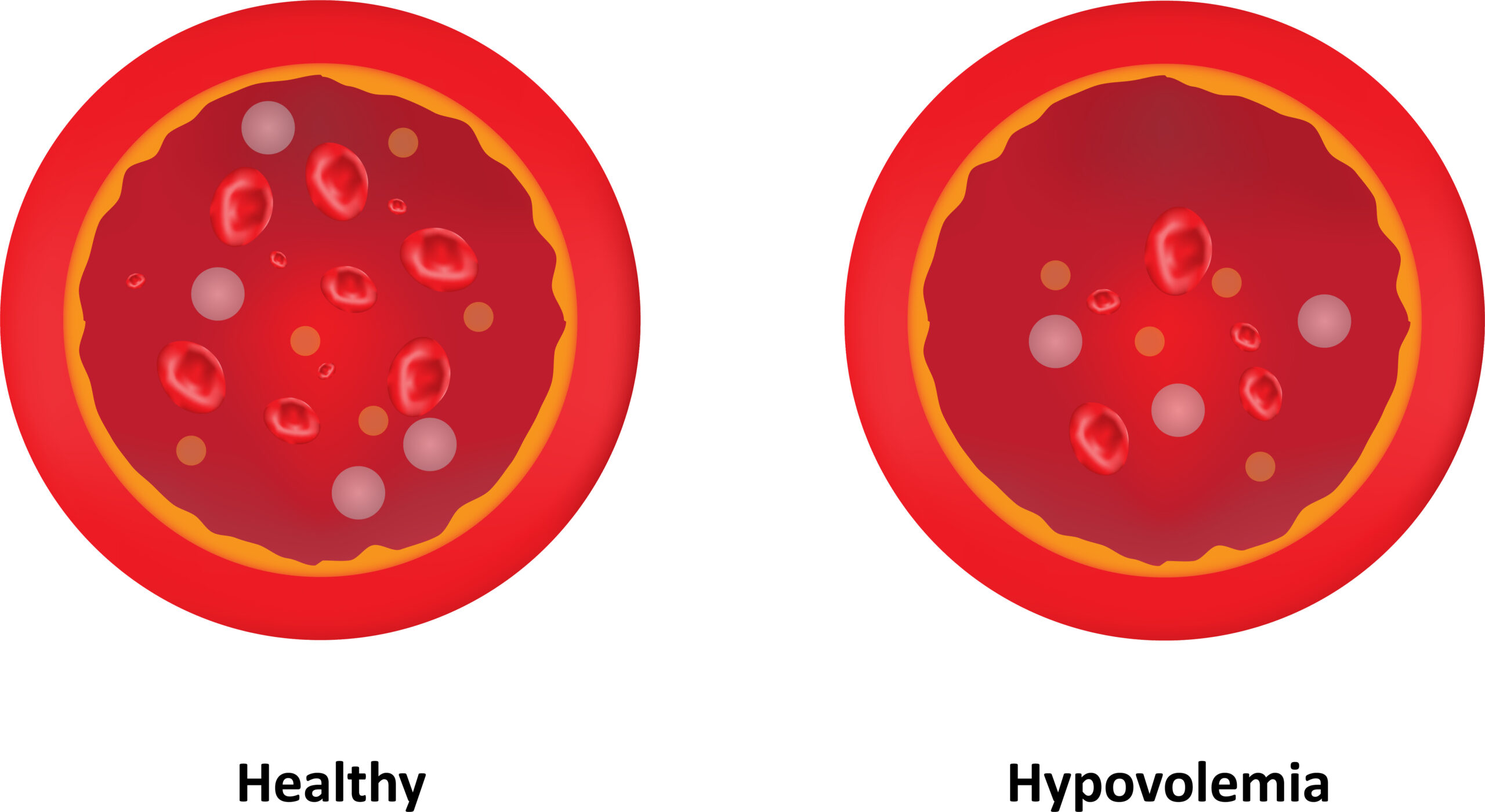
This article and video detail Shock: Cardiogenic, Hypovolemic, and Septic. Learn the three common types of shock, pathophysiology...