Asystole and Pulseless Electrical Activity Algorithm
This algorithm is for healthcare providers managing adult cardiac arrest with a non-shockable rhythm. It helps teams rapidly decide what to do when the monitor shows either asystole or pulseless electrical activity (PEA). Asystole is an absence of organized electrical activity, while PEA is organized electrical activity on the ECG without a palpable pulse. Both require immediate high-quality CPR, early epinephrine, and an aggressive search for reversible causes.
ACLS Certification Association videos have been peer-reviewed for medical accuracy by the ACA medical review board.
Article at a Glance
- PEA and asystole are nonshockable rhythms.
- Upon identification of PEA or asystole, clinicians immediately start high-quality CPR, beginning with chest compressions.
- Epinephrine is the only drug used to treat PEA and asystole.
Asystole is a cardiac arrest rhythm defined by the absence of organized ventricular electrical activity. Since there is no ventricular depolarization to reset, defibrillation is not indicated. The priorities are immediate high-quality CPR, early epinephrine, confirmation that true asystole is present, and aggressive cause correction.Asystole Algorithm (Nonshockable Rhythm)
Pulseless Electrical Activity (PEA) Algorithm
PEA is an organized electrical activity seen on ECG without a palpable pulse. Defibrillation is not indicated because the issue is not a shockable electrical circuit problem; it is a failure of mechanical output or severe physiologic collapse. The priorities are immediate CPR, early epinephrine, and rapid identification of the underlying cause.
- Confirm pulselessness: organized rhythm on monitor but no pulse, treat as cardiac arrest
- Start CPR: compressions immediately, rotate the compressor every 2 minutes
- Epinephrine: administer early, do not delay while searching for the cause
- Airway and ventilation: oxygenation, ventilation, waveform capnography when available
- Focused cause search: treat reversible causes of cardiac arrest, especially hypoxia, hypovolemia, tamponade, tension pneumothorax, thrombosis, and toxidromes
Why Asystole and PEA Are Nonshockable Rhythms
Defibrillation works by delivering an electrical shock to interrupt disorganized ventricular electrical activity, most classically ventricular fibrillation or pulseless ventricular tachycardia, so the heart can restart a coordinated rhythm. In asystole, there is no meaningful electrical activity to reset. In PEA, the electrical system may appear organized, but the problem is the absence of effective mechanical circulation. Because the core issue is not a shockable electrical rhythm, the protocol focuses on high-quality CPR, early epinephrine, minimizing interruptions, and correcting the reversible cause that prevented cardiac output.

Adult Cardiac Arrest Algorithm
Reversible Causes of PEA and Asystole (Hs and Ts)
When a patient is in pulseless electrical activity (PEA) or asystole, the best chance of return of spontaneous circulation often comes from finding and treating a reversible cause, such as cardiac tamponade. Use the Hs and Ts checklist during CPR cycles and treat what is most likely based on the clinical context.
Hs and Ts Quick Reference Table
| Category | Cause | What it means in one line |
|---|---|---|
| H | Hypovolemia | Low circulating volume reduces preload and cardiac output |
| H | Hypoxia | Inadequate oxygen delivery leads to rapid hemodynamic collapse |
| H | Hydrogen ion (acidosis) | Severe acidosis depresses myocardial function and responsiveness |
| H | Hypo or hyperkalemia | Potassium abnormalities can stop effective electrical and mechanical activity |
| H | Hypothermia | Low core temperature slows physiology and can cause arrest rhythms |
| T | Tension pneumothorax | Obstructive shock that prevents venous return to the heart |
| T | Cardiac tamponade | Pericardial pressure blocks ventricular filling |
| T | Toxins | Drug effects can cause severe bradycardia, hypotension, or arrest |
| T | Thrombosis pulmonary | Massive pulmonary embolism causes sudden obstructive collapse |
| T | Thrombosis coronary | Acute coronary occlusion can trigger arrest and refractory instability |
For a deeper explanation of each cause and what to look for during resuscitation, see Reversible Causes of Cardiac Arrest: Hs and Ts.
Treatment of Asystole and PEA
Pulseless electrical activity (PEA) and asystole are nonshockable rhythms.1 When a cardiac arrest patient presents with either, CPR is started (or continued) immediately, beginning with chest compressions. It continues for 2 minutes before another rhythm check is repeated.

Cardiac asystole is also known as cardiac flatline. There is no electrical activity in the heart.
Key Takeaway
For PEA and asystole, epinephrine should be administered as early as possible after beginning CPR.
If the patient presents with an organized rhythm after two minutes of CPR, clinicians perform a pulse check. If a pulse is felt, clinicians proceed with post-cardiac arrest care2 (go to the Post-Cardiac Arrest Care section of this course).
Healthcare clinicians must switch places after two minutes of CPR to prevent fatigue, leading to ineffective compressions.3 Clinicians should refer to mechanical and physiological parameters, such as ETCO2, to ensure high-quality CPR.
At this point, Intravenous or intraosseous access for medication and advanced airway placement with waveform capnography should be considered. Epinephrine 1 mg (1:10,000 solution) is administered. It is the only drug administered for asystole and PEA.
What is PEA?
This video explains what pulseless electrical activity looks like, why it is considered a nonshockable rhythm, and how to approach it clinically when the monitor shows organized activity but the patient has no pulse. Use this when you need a quick review of what qualifies as PEA and why cause correction matters. Watch the What is PEA video lesson.
ECG Rhythm Review: Asystole
This video reviews how asystole appears on ECG and how to avoid common false asystole errors such as loose leads or incorrect gain settings. Use this information during rhythm checks to confirm the rhythm and stay focused on CPR, epinephrine, and reversible causes. Watch the ECG Rhythm Review: Asystole lesson.
If a patient presents with a PEA or asystole, clinicians should immediately begin high-quality CPR, starting with chest compressions. Clinicians administer CPR for two minutes before checking for a pulse. The only drug clinicians should administer for asystole or PEA is epinephrine. PEA and asystole are nonshockable rhythms. Defibrillation is not indicated. Asystole and pulseless electrical activity (PEA) are both nonshockable cardiac arrest rhythms, but they are not the same. Asystole is the absence of organized ventricular electrical activity, while PEA is organized electrical activity on the monitor without a palpable pulse. In both cases, the immediate priorities are high-quality CPR with minimal interruptions, early epinephrine once IV or IO access is established, and repeated rhythm checks every 2 minutes. Defibrillation is not indicated because the problem is not a shockable rhythm. The best chance of return of spontaneous circulation comes from identifying and treating reversible causes using the Hs and Ts during CPR cycles, while maintaining effective compressions and ventilation support.Summary
More Free Resources to Keep You at Your Best
Editorial Sources
ACLS Certification Association (ACA) uses only high-quality medical resources and peer-reviewed studies to support the facts within our articles. Explore our editorial process to learn how our content reflects clinical accuracy and the latest best practices in medicine. As an ACA Authorized Training Center, all content is reviewed for medical accuracy by the ACA Medical Review Board.
1. Tony I. Oliver; Usama Sadiq; Shamai A. Grossman. Pulseless Electrical Activity. National Library of Medicine. 2022.
2. American Heart Association. Adult Basic Life Support. 2010.
3. Jesse Borke, MD, FACEP, FAAEM; Chief Editor: Kirsten A Bechtel, MD. Cardiopulmonary Resuscitation (CPR). Medscape. 2021.
More to Learn
Fibrinolytic and endovascular therapies are the immediate therapies involved in stroke treatment. Read our detailed article on immediate therapies and their inclusion/exclusion criteria.
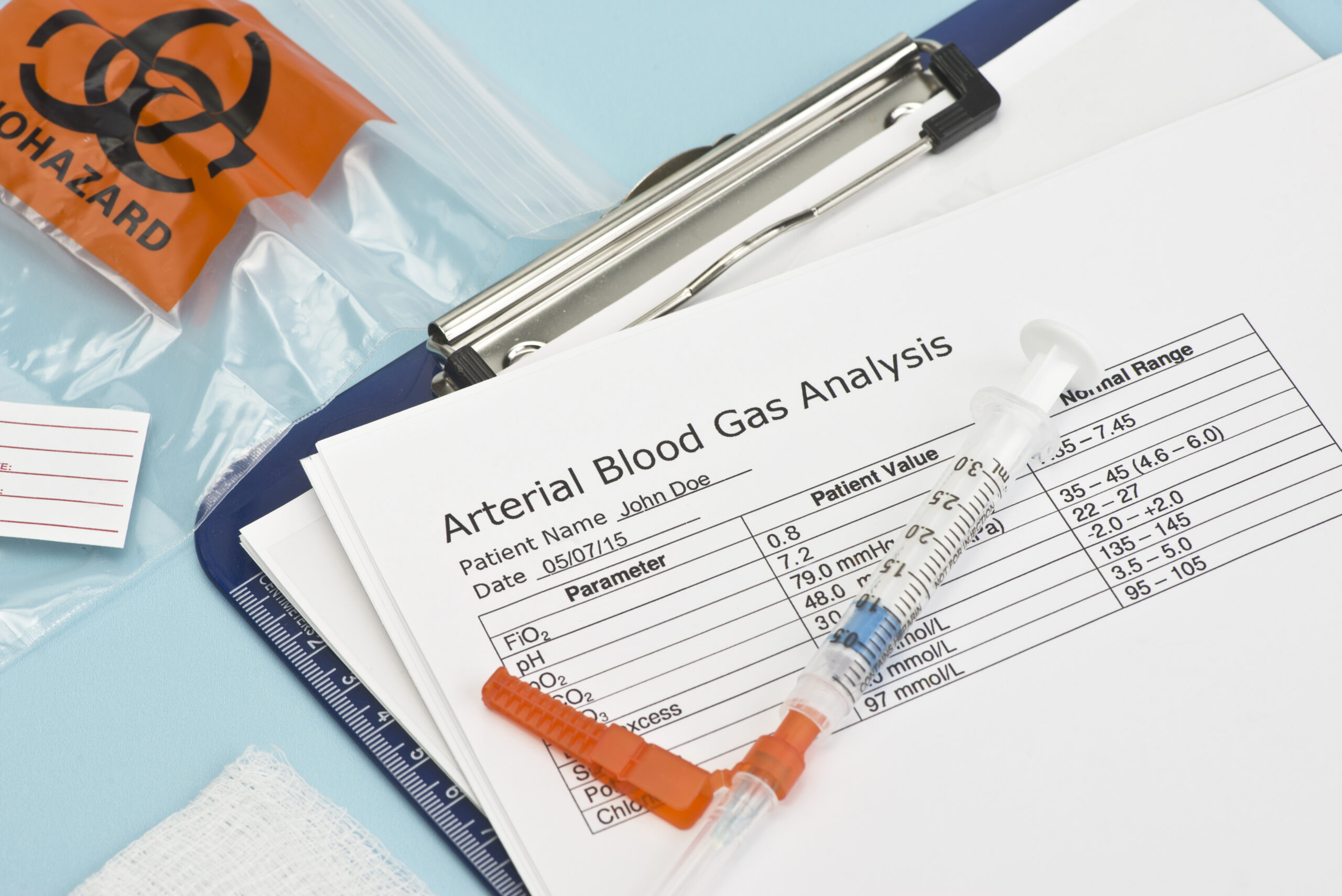
This article and video show An Easy Way to Interpret Arterial Blood Gases. Learn how to interpret arterial blood gases...