ACLS Hs & Ts – Cardiac Tamponade
ACLS Certification Association videos have been peer-reviewed for medical accuracy by the ACA medical review board.
Article at a Glance
- Cardiac tamponade is one of the Hs and Ts and a reversible cause of cardiac arrest.
- When fluid accumulates in the pericardial sac, cardiac tamponade can result.
- Cardiac tamponade prevents the heart from filling during diastole.
- Symptoms include muffled heart tones, JVD, and hypotension – Beck’s Triad.
- Treatment includes an infusion of normal saline and removal of fluid from the pericardial sac.
Cardiac tamponade occurs in two ways, acute and subacute. To understand these better, we must review the anatomy of the pericardial sac (also known as pericardium).Cardiac Tamponade
Anatomy of the Pericardial Sac
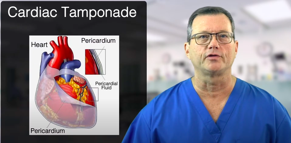
The pericardium is a sac that covers the heart.
The pericardial sac’s outside layer is fibrous and tough. There are two more inner layers, the parietal layer and the visceral layer. The visceral layer is closest to the heart.
Pericardial fluid exists between the parietal and visceral layers, and there is usually around 50 mL of fluid in the pericardial sac. The fluid acts as a lubricant, so the heart can beat without rubbing itself raw.
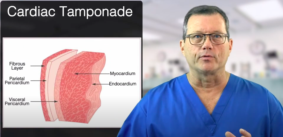
The pericardium consists of the fibrous layer, parietal layer, and visceral layer.
Acute Cardiac Tamponade
Acute means something that rapidly occurred. Most acute cardiac tamponades are associated with trauma, such as a blunt force to the chest. For example, the heart is lacerated, and the bleeding starts to fill up the pericardial sac. The heart constricts because the pericardial sac doesn’t stretch very quickly. That prevents the heart from filling up during diastole, causing near diastolic failure.
Treatment
Physicians treat acute cardiac tamponade by relieving the blood from the pericardial sac. A physician in the emergency department (ED) will use a needle and tap the pericardial sac to remove the fluid.
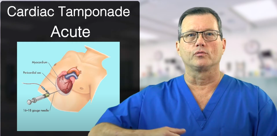
Pericardial Sac Fluid Removal Procedure
Beck’s Triad
It’s essential to recognize acute cardiac tamponade in the field. The three collective symptoms are known as Beck’s Triad. They are:
- Muffled heart tones
- Hypotension
- Jugular vein distension
Muffled heart tones are characterized by a decreased heart tone intensity, as though the heart tones are dampened. Physicians must know a regular heart tone to diagnose one that’s muffled.
Next is hypotension. Blood pressure drops because the blood cannot get back into the heart.
Lastly, is jugular vein distention (JVD). The blood cannot load back into the heart, so it backs up in the venous system.
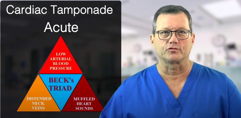
The three symptoms of cardiac tamponade are known as Beck’s Triad.
Subacute Pericardial Tamponade
Subacute pericardial tamponades develop in a few ways. It may occur in the presence of bleeding directly from the heart, such as during a catheterization procedure where micro-lacerations in the vessels are slowly leaking. Studies show that this happens 0.1–0.6% of the time. You can detect micro-lacerations by performing an echocardiogram two weeks after the patient’s catheterization lab visit.
Pleural effusion is another cause. In this case, the heart is not bleeding. The main problem is an increased amount of pericardial fluid between the two layers of the pericardial sac.
Pleural effusion usually happens or is associated with pericarditis, an infection of the heart leading to an increased amount of fluid within the pericardial sac. Between the parietal and the visceral layers, fluid starts to build up secondary to an infection.
Related Video – What is Pleural Effusion?
There are instances where a physician has tapped and removed almost two liters of fluid due to a pleural effusion. The chest X-rays in the image below display a huge heart. If we look at a CT scan, we will clearly see the pericardial sac filling with fluid.
Read: Reversible Causes of Cardiac Arrest: Hs and Ts
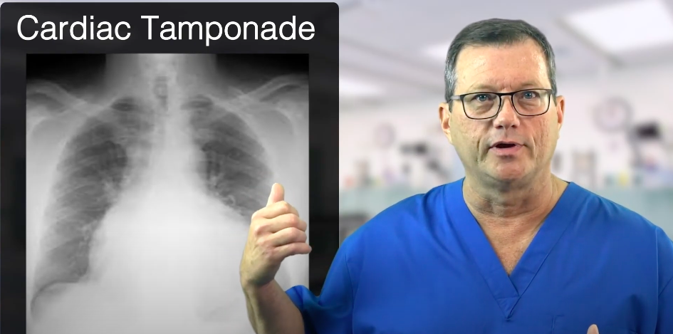
An Enlarged Heart Due to Pleural Effusion.
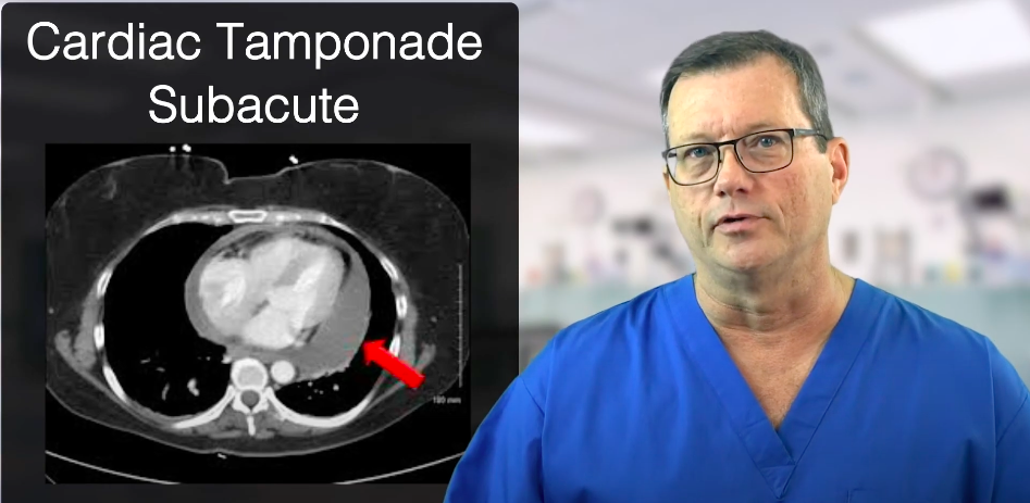
A CT scan of cardiac tamponade with an arrow indicating fluid build-up in the pericardial sac.
Treatment
To treat acute symptoms of cardiac tamponade in the field, perform rapid, aggressive fluid infusion. Start with 0.9% normal saline into the patient to try to keep more fluid inside the ventricles than leaking outside of the heart, thereby compressing the heart. It’s only a temporary measure which will hopefully buy more time to reach the ED.
Summary
There are acute and subacute cardiac tamponades. Acute cardiac tamponades occur quickly, for example, blunt force trauma to the chest. The symptoms are muffled heart tones, hypotension, and jugular vein distension, collectively known as Beck’s Triad. Subacute tamponades may occur when there’s bleeding directly into the heart or a pleural effusion. Fluid infusion is the first treatment.
More Free Resources to Keep You at Your Best
Editorial Note
ACLS Certification Association (ACA) uses only high-quality medical resources and peer-reviewed studies to support the facts within our articles. Explore our editorial process to learn how our content reflects clinical accuracy and the latest best practices in medicine. As an ACA Authorized Training Center, all content is reviewed for medical accuracy by the ACA Medical Review Board.
More to Learn

QRS-complexes less than 0.12 seconds are considered narrow. Read the detailed article to learn the types of, and treatment for, Narrow QRS-Complex Tachycardia.
![]()
Learn about premature junctional contractions (PJCs) and their criteria in our article. Understand these premature beats that arise from the atrioventricular (AV) junction of the heart.