Atrial Rhythms: Rhythms Originating from the Atrium (ECG/EKG Interpretation)
Atrial rhythms are abnormal electrical patterns originating from the upper chambers of the heart (the atria) rather than the primary pacemaker. Designed for nursing and medical professionals, this guide will help you confidently identify these rhythms on an ECG using a reliable, repeatable checklist.
ACLS Certification Association videos have been peer-reviewed for medical accuracy by the ACA medical review board.
Article at a Glance
- Several abnormal rhythms are caused by foci conducting impulses from areas other than the sinoatrial (SA) node.
- Rhythms originating outside the sinoatrial node but within the atria are recognizable due to P wave size and shape.
- Rhythms originating from the atria are termed “supraventricular” because they originate above the ventricles, which also generally results in a narrow QRS complex.
SA Node Irregularity
When the SA node fails to generate an impulse, a site outside the SA node but within the atria will generate an action potential faster than the SA node. The heart switches to the next fastest inherent rate beyond the SA node.
If the SA node isn’t functioning, the atrial focus initially takes over, becoming the predominant pacemaker. The atria generate impulses which travel towards the Purkinje fibers (the network of specialized cells that cause the ventricles to contract) via the atrioventricular (AV) junction (the electrical bridge between the atria and ventricles) before heading to the ventricles the same way they would if the signal came from the SA node.
If the rate is faster than 100 beats per minute (bpm), the term “atrial tachycardia” describes the rhythm. If the rate is less than 100 bpm, it’s an “ectopic atrial” rhythm.
Impulses originating in the atrium change the normal morphology (shape) of the P wave. The atrial P wave is slower than the sinus P wave, and its morphology may be flattened, notched, or peaked. In certain specific rhythms like atrial flutter, it may have a saw-tooth appearance, while others may present a biphasic appearance.
A biphasic wave may produce a positive (upward) and negative (downward) deflection from the isoelectric line (the flat baseline of the ECG tracing) within one P wave. Rhythms originating from the atria will generally have a narrow QRS complex because the impulses are generated above the ventricles.
Clinical Callout: Morphology Assessment
When assessing these P waves, always ask yourself: “Do they all look normal?” Changes in size, shape, or direction are key indicators of an atrial origin.
Wandering Pacemaker
A wandering pacemaker has different foci of impulse generation throughout the atrium, switching between the sinoatrial node, atrium, and atrioventricular junction. The resulting rhythm is a combination of sinus and atrial beats.
An impulse originating from the sinoatrial node presents a smooth, rounded P wave with an upward deflection. An impulse originating from the atrium presents a flattened, notched, or biphasic morphology. An impulse originating from the atrioventricular junction will result in a P wave which may look inverted or may be absent.
The PR interval also varies as the impulse is generated from different foci.

P wave morphology changes from one complex to the next on a wandering pacemaker’s ECG strip.
- RATE: normal rate is between 60–100 bpm
- RHYTHM: varying R-R intervals due to changes in pacemaker sites
- P WAVE: morphology changes from one complex to the next. The P wave is followed by the QRS complex
- PR INTERVAL: varies but less than 0.20 seconds
- QRS COMPLEX: less than 0.12 seconds
Ectopic Beat
An ectopic beat is a single beat from a focus outside the SA node but within the conduction system.1 Rather than being classified as a sustained arrhythmia on its own, it often represents an irritability in the conduction system, generating an impulse strong enough to override the impulse generated from the SA node. An ectopic beat may also originate as an impulse from an escape mechanism. It is important to distinguish between a single ectopic beat interrupting a normal heart rhythm and a sustained ectopic atrial rhythm. An ectopic beat arising from the atria is known as an atrial ectopic.
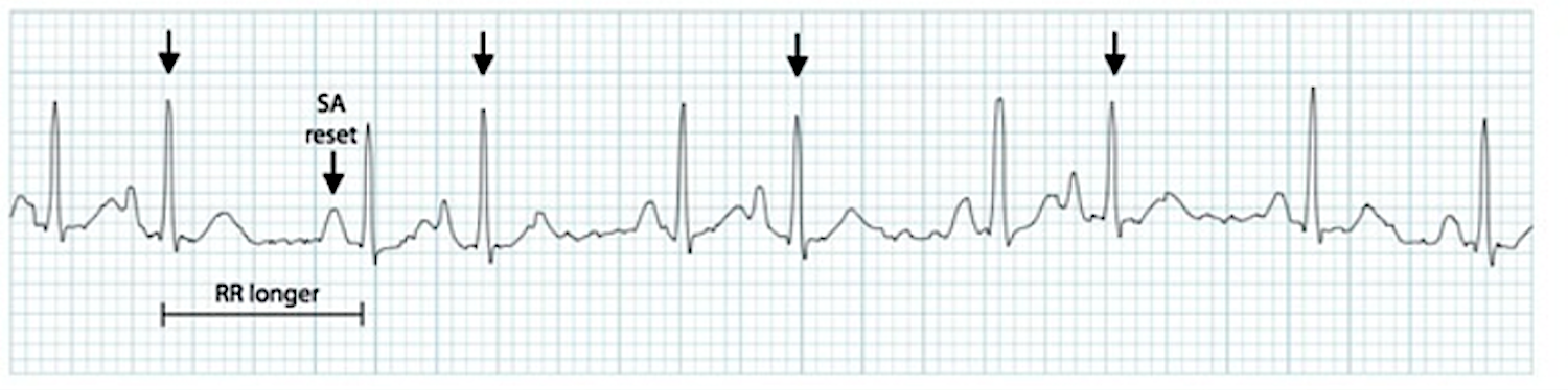
When irritability in the conduction system causes a beat to occur from an area in the atria other than the SA node, it’s a premature atrial contraction (PAC). ECG strip detailing a premature atrial contraction (PAC). The video below provides a visual breakdown of Premature Atrial Contractions (PACs). Watching this will help you understand how an early beat interrupts the underlying rhythm and how to spot it on an ECG tracing. PACs usually occur as a single beat. Clinicians should report the core or underlying rhythm and the type of ectopic beat (i.e., normal sinus rhythm with PAC) when reporting an ECG interpretation with a premature atrial contraction. A PAC may make the rhythm appear irregular, yet the clinician still measures R-R intervals on the 6-second strip to determine if the core rhythm is regular or irregular. The above strip details several PACs and the rhythm is quite irregular. PACs may appear in patterns, such as every other beat (bigeminy) or every third beat (trigeminy). It is also possible a PAC occurs near the end of the QRS complex and the P wave, manifesting near the T wave. It may be entirely lost within the T wave. The below strip illustrates how the T wave appears more peaked than others.Premature Atrial Contraction
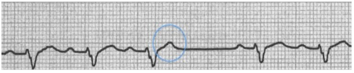
A PAC presents with a peaked T wave on an ECG.
- RATE: depends on the core rhythm
- RHYTHM: depends on the core rhythm, which is interrupted when the PAC occurs
- P WAVE: flattened, notched, peaked or biphasic; it may occur near, or be buried within, the T wave
- PR INTERVAL: 0.12–0.20 seconds or longer
- QRS COMPLEX: less than 0.12 seconds
Atrial Tachycardia
An irritable focus within the atria causes atrial tachycardia.3 The impulse generated is greater than the impulse from the SA node, the normal pacemaker of the heart. Individual complexes in atrial tachycardia have the characteristics of a PAC, including P wave morphology, PR-interval, and QRS complex.
However, atrial tachycardia involves every beat instead of a single beat. While often grouped together, atrial flutter and multifocal atrial tachycardia (MAT) are closely related supraventricular tachycardias rather than strict subtypes of simple atrial tachycardia.

An ECG strip illustrating multifocal atrial tachycardia (MAT) with impulses originating in multiple foci within the atria. The arrows point to P waves with unusual morphology.
Atrial tachycardia generally presents as a regular arrhythmia with heart rates commonly ranging from 150 to 250 bpm, though associated atrial rhythms can reach higher rates.
- RATE: typically 150–250 bpm
- RHYTHM: regular (may be irregular in MAT)
- P WAVE: atrial P wave is different in morphology from a sinus P wave. The P wave may be buried within a T wave.
- PR INTERVAL: 0.12–0.20 seconds
- QRS COMPLEX: less than 0.12 seconds
ECG Rhythm Review – Atrial Tachycardia
Watch the following video for an in-depth rhythm review of Atrial Tachycardia. This resource will help you identify the rapid rate and altered P wave morphology characteristic of this rhythm.
Read: Rhythms Originating from The Sinus Node
When the atria are intensely irritable, a cardiac rate above 250–350 bpm may occur. The irritability causes a rhythm known as atrial flutter, represented by a saw-tooth appearance of the P waves due to the cyclic pattern of generated impulses creating a series of rapid atrial waves. An anti-clockwise pattern of impulse generation reveals a negative deflection of the P wave on the LEAD II. If the impulse generated flows in an anti-clockwise manner, LEAD II shows a negative deflection of P waves. When the rapid rate occurs, the ventricles do not have enough time to adequately fill, so there is not enough blood volume ejected to sufficiently meet the body’s needs. Fortunately, the atrioventricular node (AV node) regulates the heart, blocking some impulses from reaching the ventricles. That allows the ventricles to fill for adequate ejection into the systemic circulation. The AV node allows impulses to pass through every second, third, or fourth P wave.4 A 12-lead ECG will detail a rapid succession of P waves with a rate between 250 and 350 bpm. A QRS complex does not follow every P wave, only manifesting every second, third, or fourth saw-tooth wave. That causes a slower ventricular rate compared to the atrial rate. The QRS complex only manifests every two, three, or four saw-tooth waves, creating a 2:1, 3:1, or 4:1 block. If the R-R interval is irregular, variable blocks are occurring.Atrial Flutter
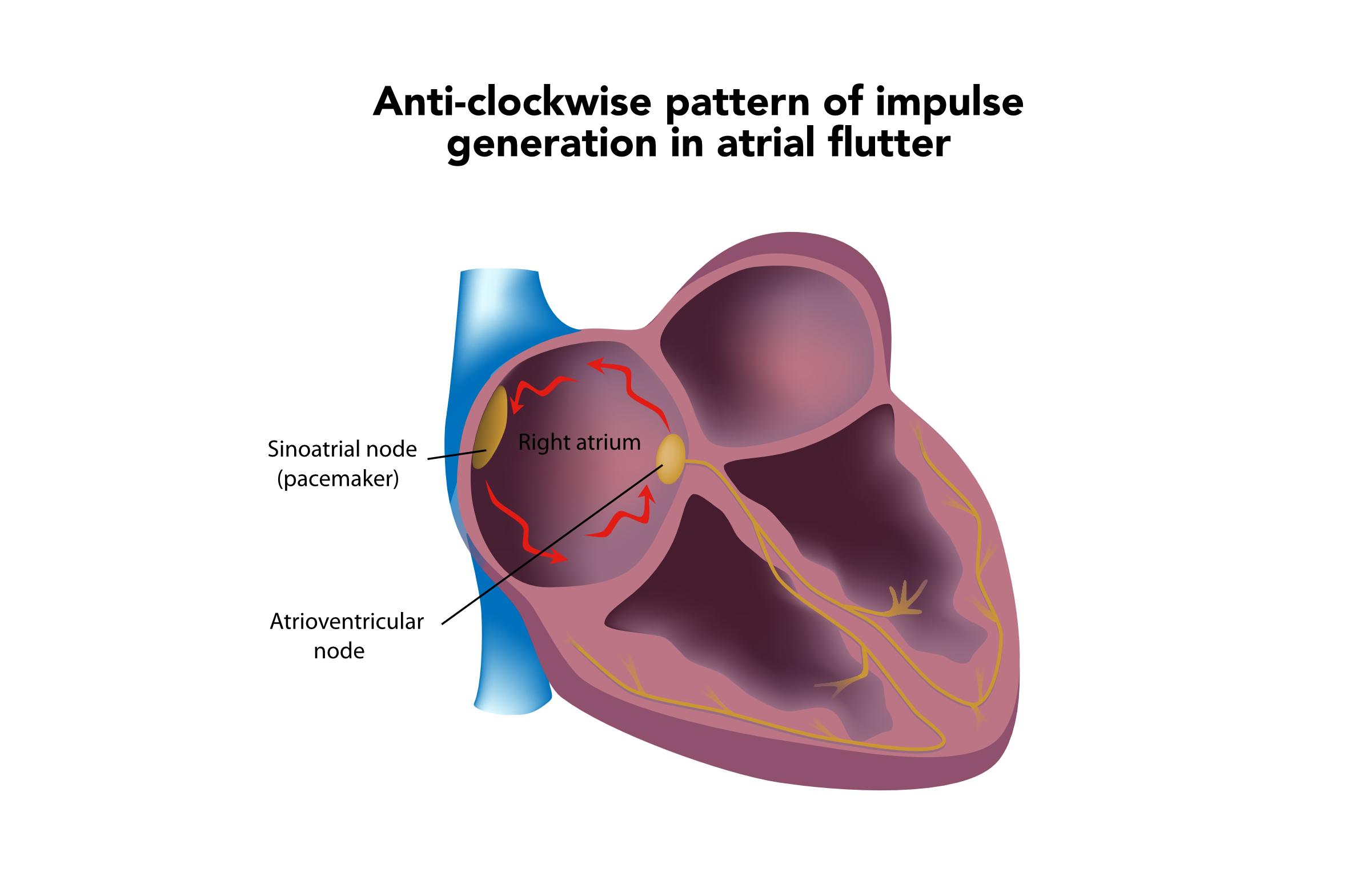

A 3:1 block with an atrial rate of 250 bpm and a ventricular rate of 65 bpm.
In atrial flutter, the PR interval is not measured because it cannot be determined from the ECG strip. The QRS complex is normal at 0.12 seconds.
- RATE: atrial rate of 250–350 bpm; ventricular rate varies
- RHYTHM: atrial rhythm is regular; ventricular rhythm is usually regular but may be irregular if a variable block is present
- P WAVE: saw-tooth appearance (specifically referred to as flutter waves)
- PR INTERVAL: cannot be determined
- QRS COMPLEX: less than 0.12 seconds
Pitfall: It can be easy to confuse atrial flutter with variable block for atrial fibrillation because both can present with an irregular R-R interval. However, atrial flutter will present with distinct, regular saw-tooth flutter waves, whereas atrial fibrillation features chaotic, disorganized fibrillatory waves.
ECG Rhythm Review – Atrial Flutter
This video reviews the ECG criteria for Atrial Flutter. It demonstrates how to spot the classic saw-tooth pattern and calculate the conduction block ratio.
Atrial Fibrillation
During atrial fibrillation, the atria are so intensely irritable they no longer beat but merely quiver.5 The ECG tracing displays barely visible fibrillatory waves within the isoelectric line (the flat baseline of the ECG tracing), known as F waves. The quivering atria generate an impulse up to 350 bpm.
P waves are typically not discernible in atrial fibrillation. The ventricular rate is produced by the AV node blocking most of the generated impulses, producing an irregular R-R interval pattern. The F waves are generated in such a chaotic manner no discernable pattern is produced.
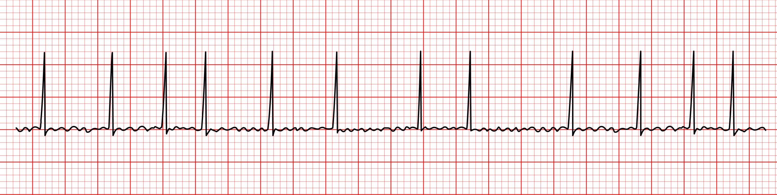
In atrial fibrillation, the R-R interval pattern is irregular.
Atrial fibrillation is easily recognized. The primary characteristic findings are the absence of visible P waves and irregular rhythms resulting from irregular R-R intervals. As the ventricular rate increases, R-R intervals may become closer and appear to have a regular rhythm.
A rapid ventricular rate above 100 bpm (or uncontrolled ventricular response) may induce symptoms in the patient, while a ventricular rate lower than 100 bpm (or controlled ventricular response) is less likely to cause symptoms. Atrial fibrillation symptoms result from the quivering atria’s inability to fill with adequate blood, resulting in reduced cardiac output.
ECG Rhythm Review – Atrial Fibrillation
Watch this video to learn how to identify Atrial Fibrillation on an ECG. It highlights the chaotic baseline and the irregularly irregular R-R intervals that are hallmarks of this common arrhythmia.
Supraventricular Tachycardia
Supraventricular tachycardia (sometimes referred to as paroxysmal supraventricular tachycardia, or PSVT) is a regular tachyarrhythmia where P waves are often not visible or may be buried, arising from either the atria or AV node.6 The rate is usually 150–250 bpm.
Due to the rhythm’s rapidity, patients are often symptomatic due to decreased cardiac output. The rhythm is rapid, regular, with P waves that are typically obscured, and is easily recognized. The QRS complex is narrow as the rhythm arises above the ventricles.

Supraventricular tachycardia features P waves that are often buried or not visible.
- RATE: 150–250 bpm
- RHYTHM: regular
- P WAVE: often not visible or buried
- PR INTERVAL: cannot be measured
- QRS COMPLEX: less than 0.12 seconds.
One Quick Question: What are the SVT (Supraventricular Tachycardia) Criteria?
This brief video reviews the definitive ECG criteria for Supraventricular Tachycardia (SVT). It focuses on identifying the rapid rate, regular rhythm, and narrow QRS complex.
What are the Differences Between ST (sinus tachycardia) and SVT?
Differentiating between Sinus Tachycardia (ST) and SVT is critical for proper treatment. This video outlines the key differences in rate, P wave presence, and onset/termination patterns.
ECG Rhythm Review – Supraventricular Tachycardia (SVT)
For a more comprehensive look, this video provides a detailed ECG rhythm review of SVT, solidifying your ability to identify it in clinical scenarios.
Frequently Asked Questions
Do they all have QRS complexes after them?
Generally, yes, because the electrical impulse travels down through the AV node to the ventricles. However, in rhythms like atrial flutter with a block (e.g., 2:1, 3:1), the AV node protects the ventricles from the rapid atrial rate, meaning not every atrial impulse results in a QRS complex.
Do they all have P waves in front of them?
Not necessarily. While many atrial rhythms display visible (though morphologically abnormal) P waves, rhythms like atrial fibrillation feature chaotic fibrillatory (F) waves instead of true P waves. Additionally, in rapid rhythms like SVT, the P wave is often buried in the preceding T wave and is not visible on the ECG.
Summary
Abnormal rhythms originating in the atrium occur when foci outside the SA node dictate the heart’s electrical activity. To accurately diagnose and treat these supraventricular rhythms, such as wandering pacemakers, premature atrial contractions (PACs), atrial flutter, atrial fibrillation, and SVT, clinicians must systematically evaluate the ECG using a consistent criteria framework:
- Rate: Note if the atrial and ventricular rates are normal, bradycardic, or tachycardic.
- Regularity (Rhythm): Determine if the R-R intervals are regular, irregularly irregular (like AFib), or interrupted by early beats (like PACs).
- P Waves: Assess morphology, are they flattened, notched, peaked, biphasic, saw-tooth, fibrillatory, or buried?
- PR Interval: Measure to see if it is consistent, variable, or unmeasurable.
- QRS Complex: Confirm it is narrow (less than 0.12 seconds), characteristic of supraventricular origins.
More Free Resources to Keep You at Your Best
Editorial Sources
ACLS Certification Association (ACA) uses only high-quality medical resources and peer-reviewed studies to support the facts within our articles. Explore our editorial process to learn how our content reflects clinical accuracy and the latest best practices in medicine. As an ACA Authorized Training Center, all content is reviewed for medical accuracy by the ACA Medical Review Board.
1. Markham Heid. What Is an Ectopic Beat? WebMD. 2022.
2. Joseph Heaton; Srikanth Yandrapalli. Premature Atrial Contractions. National Library of Medicine. 2021.
3. Johns Hopkins Medicine. Atrial Tachycardia.
4. Joseph Heaton; Amandeep Goyal. Atrioventricular Node. National Library of Medicine. 2021.
5. Rachel Nall, MSN, CRNA. Improving Your Atrial Fibrillation Prognosis. healthline. 2021.
6. Laryssa Patti; John V. Ashurst. Supraventricular Tachycardia. National Library of Medicine. 2022.
More to Learn
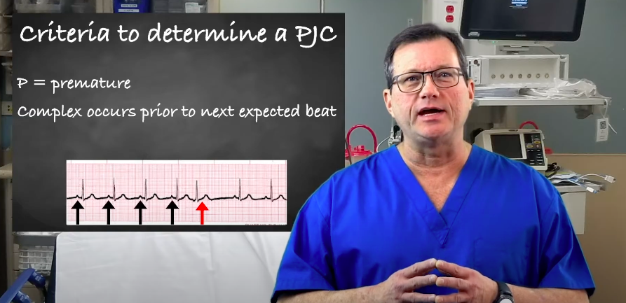
Learn about premature junctional contractions (PJCs) and their criteria in our article. Understand these premature beats that arise from the atrioventricular (AV) junction of the heart.
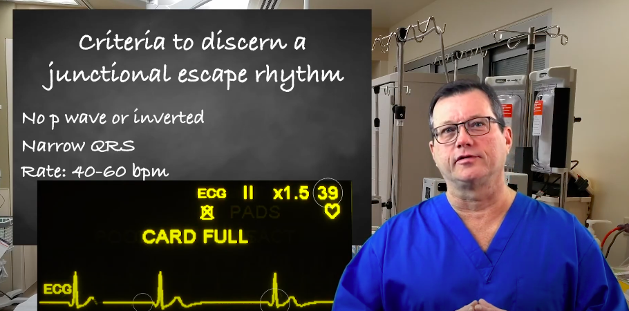
Learn about junctional escape rhythm criteria for ACLS certification with our article. Understand the guidelines and requirements seamlessly.