Normal ECG Variants Flashcard
Rationale
A. Rationale: An ectopic beat is produced by an irritable focus strong enough to overcome the sinoatrial node impulse within the conduction system. It can also be generated as a form of escape mechanism.
Question
A group of rhythms originating outside of the sinoatrial node but still within the conduction system that represents an IRRITABILITY in the conduction system is known as which of the following arrhythmias?
a. Ectopic beat
b. Atrial fibrillation
c. Ventricular fibrillation
d. Wandering pacemaker
Answer
a. Ectopic beat
Rationale
D. Rationale: The description above points to a sinus rhythm except for the slow heart rate < 60 bpm. Therefore, this describes sinus bradycardia. AV blocks have delayed impulses traveling from the atria to the ventricles; hence, they should present with prolonged PR interval.
Question
An ECG tracing represents a heart rate of 48 bpm, regular R-R interval, an upward deflection of the P wave, an R-R interval of 0.12 seconds, and a QRS complex < 0.12 seconds. What ECG pattern does this describe?
a. First-degree AV block
b. Second-degree Mobitz type I AV block
c. Complete AV block
d. Sinus bradycardia
Answer
d. Sinus bradycardia
Rationale
D. Rationale: Junctional P waves can occur before the QRS complex when it depolarizes before ventricular depolarization, they can be buried within the QRS complex when it depolarizes at the same time as ventricular depolarization, and they may occur after the QRS complex when ventricular depolarization occurs before the junctional depolarization.
Question
Patients with a junctional rhythm have junctional P waves that may occur in which segment of the ECG tracing?
a. Before the QRS complex
b. After the QRS complex
c. Buried within the QRS complex
d. All of the above describe junctional P waves
Answer
d. All of the above describe junctional P waves
Rationale
C. Rationale: An old myocardial infarction presents with Q waves and symmetric negative T waves concurrent with a medical history or increased risk of coronary artery disease.
Answer choice A – Myocardial ischemia shows ST depression on ECG leads with the corresponding left ventricular wall.
Answer choice B – Severe hypokalemia is indicated by T wave inversion and prominent U waves.
Answer choice D – A bundle branch block pattern shows an rsR’ pattern.
Question
When analyzing a patient’s ECG, you notice a deep Q wave in frontal lead II and inverted T wave morphology in frontal lead III. What can you conclude?
a. Myocardial ischemia
b. Severe hypokalemia
c. Old myocardial infarction
d. Bundle branch block
Answer
c. Old myocardial infarction
Rationale
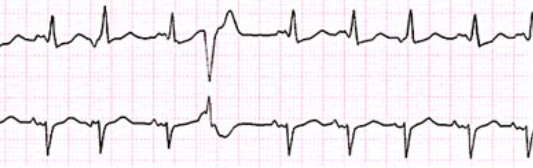
C. Rationale: The R on T phenomenon is represented by an R wave of the PVC that overlaps with the T wave of the core rhythm due to a strong ventricular depolarization that has caused it to have a relative refractory state. The charges that are still polarized are emitted during the PVC event.
Question
Which characteristic of a premature ventricular contraction is caused by a refractory state of the T wave of the core rhythm when an irritable ventricular focus causes a strong ventricular depolarization?
a. Premature ventricular contraction
b. ST elevation
c. R on T phenomenon
d. Torsade’s de pointes
Answer
c. R on T phenomenon
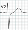
Rationale
C. Rationale: The ECG in choice C shows a pathologic Q wave with an inverted T wave, which is a hallmark finding for patients with clinical presentations of myocardial infarction.
Answer choices A, B, and D – A pathologic Q wave is wider than 40 ms.
Question
Which one of the following ECG examples is likely a pathologic Q wave?
a.

b.

c.

d.
Answer
c.
Rationale
C. Rationale: Frontal lead III and sometimes frontal lead aVF may have a deep Q wave that is non pathologic. This diagnosis is confirmed when the patient has no clinical findings or symptoms that represent coronary heart disease.
Answer choices A, B, and D – Q waves are considered pathologic and may represent myocardial infarction, systolic overload, left ventricular hypertrophy, or a left bundle-branch block.
Question
Which one of the following frontal leads may have non pathologic or normal variant deep Q waves?
a. Lead I
b. Lead II
c. Lead III
d. Lead aVL
Answer
c. Lead III
Rationale
B. Rationale: The normal PR interval is 0.12–0.20 seconds. This helps to determine if every P wave is conducted. If not all P waves are conducted, there might be a second-degree AV block.
Answer choice A – If an impulse is generated from the ventricles, there generally will be no P waves present on ECG.
Answer choice C – Lack of conduction of P waves is always an abnormal ECG finding.
Answer choice D – In atrial fibrillation, there will be fibrillatory waves present, as opposed to P waves.
Question
While measuring the PR interval, you notice that not all P waves are conducted throughout the long lead ECG tracing. What is the most probable conclusion?
a. The impulse may be generated from the ventricles.
b. There could be some form of second-degree AV block.
c. This could be a normal variant.
d. The patient may be in atrial fibrillation.
Answer
b. There could be some form of second-degree AV block.