Chest Tubes: Indications, Components, Nursing Assessments, and Care
ACLS Certification Association videos have been peer-reviewed for medical accuracy by the ACA medical review board.
Table of Contents
- Purpose of Chest Tubes
- Parts of the Chest Tube
- Tubing
- Drainage Holes
- Collection Chamber
- Water Seal Chamber
- Suction Regulator
- Suction Monitor Bellows
- Nursing Assessments and Care
- Water Seal
- Character of Output
- Amount of Output
- Bubbling
- Tidaling
- Relative Location to the Chest
- Avoid Milking the Tube
- Petroleum Gauze and Tape
- Sterile Water
- Clamping
Article at a Glance
- A chest tube is used to drain fluid, blood, or air from the chest.
- The chest tube drainage system contains several parts, each with a specific purpose.
- Read on to learn about the nursing assessments and care of chest tubes.
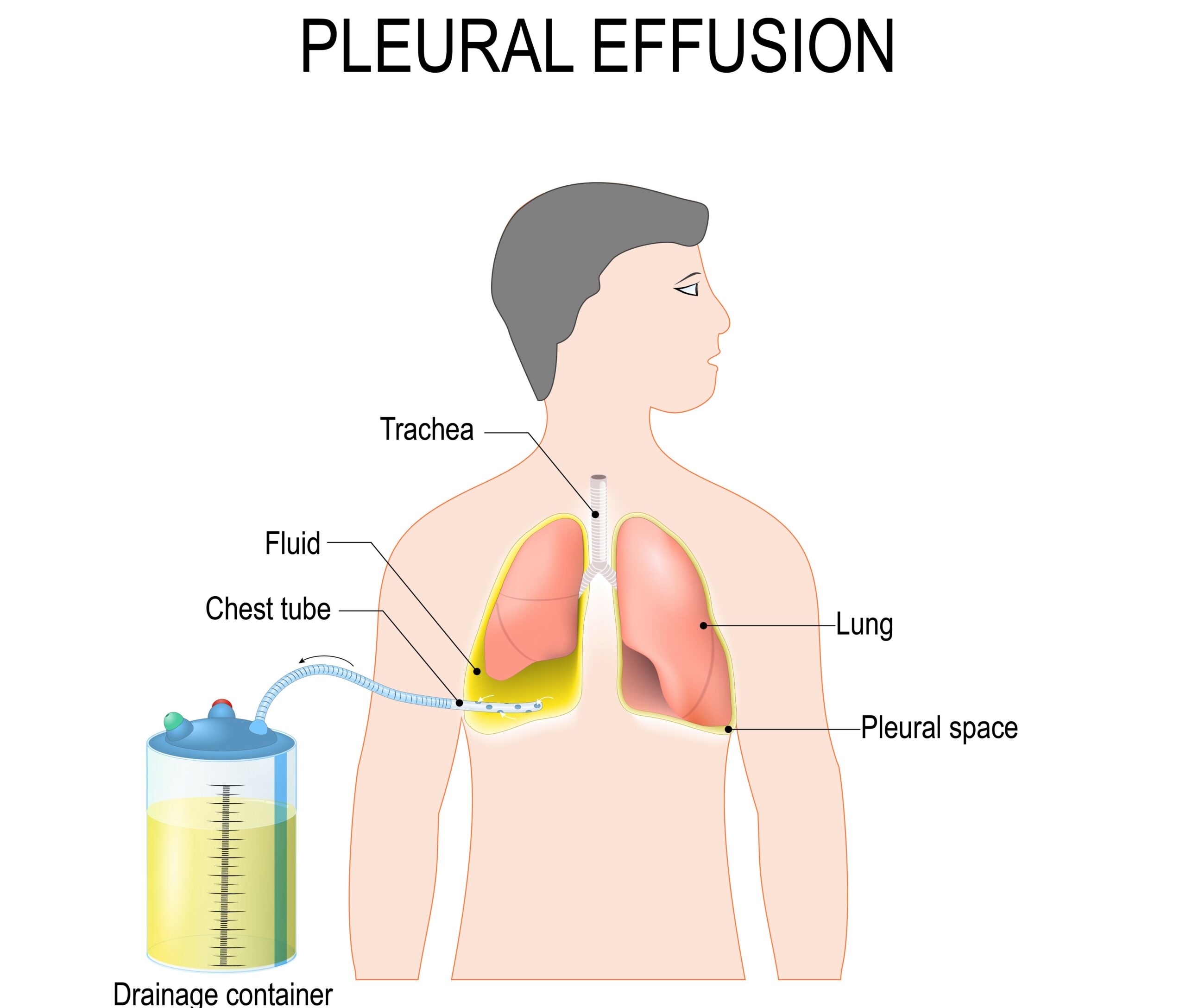
This article introduces chest tubes, including their uses, parts, and care. A chest tube is a hollow tube placed into the chest to drain fluid, blood, or air. For the most part, chest tubes are inserted to facilitate lung expansion. When a patient has pneumothorax or pleural effusion, there is either air or fluid build-up within the pleural space. That fluid prevents full lung expansion. In addition to facilitating lung expansion, mediastinum chest tubes are also sometimes inserted in the sternal area after a cardiovascular surgery. The mediastinum chest tube prevents cardiac tamponade, which occurs if fluid builds up around the area of incision after cardiovascular surgery. The chest tube helps to keep fluid off the heart and facilitate healing. A pneumothorax is a collapsed lung due to air leaking into the pleural space. A chest tube can be used to remove air from the pleural space.Purpose of Chest Tubes
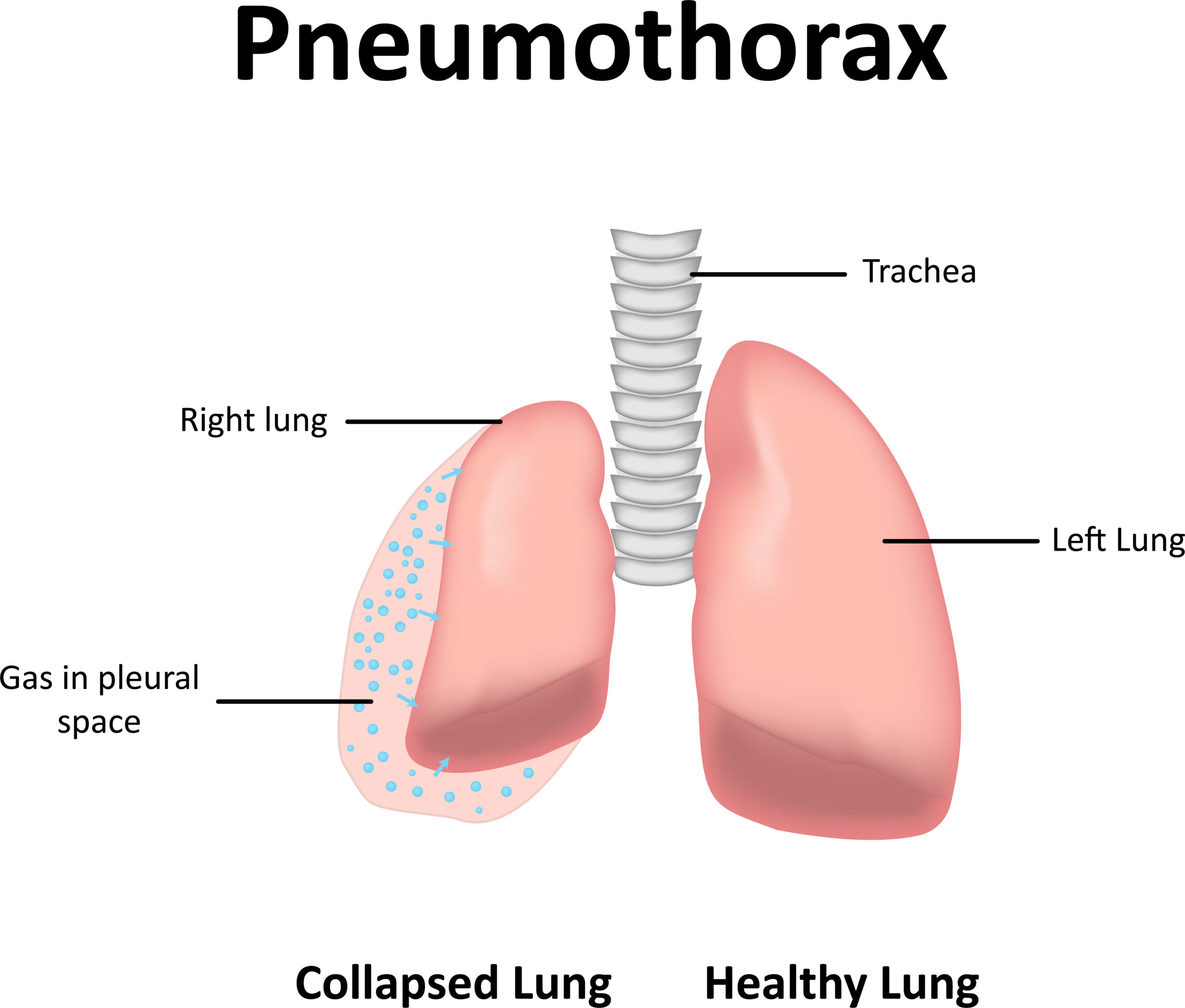
Related Video – What is Pneumothorax?
The main parts of the chest tube are:Parts of the Chest Tube
The provider will choose the size of the chest tube depending on what needs to be drained. Generally, chest tubes are large and painful upon insertion.Tubing
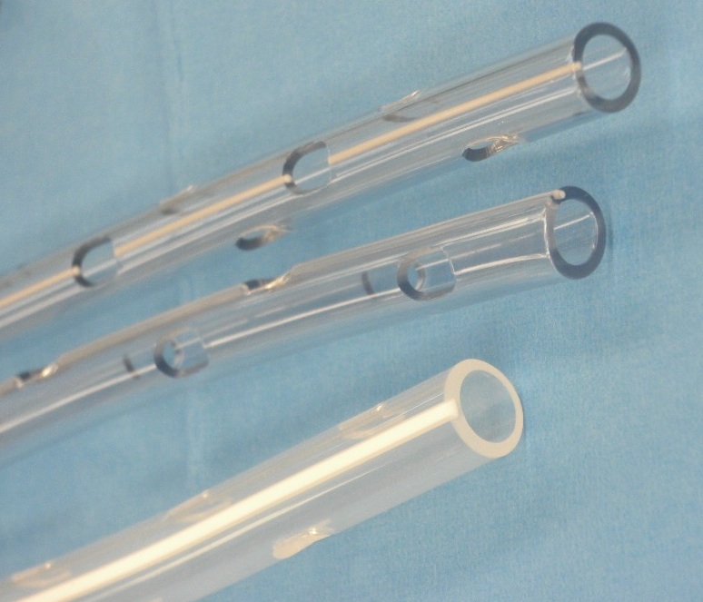
At the tip of the tube are small holes called drainage holes. These holes allow air or fluid to enter the tube. Caption: Chest tube drainage holes allow fluid or air to flow into the tubing.Drainage Holes
The collection chamber collects what is being drained from the chest tube. Typically, the collection chamber collects fluid. Whatever drains into the collection chamber is documented in the intakes and outputs. Of note, if a patient has a pneumothorax, not much will appear in the collection chamber because the tube is removing air.Collection Chamber
The water seal chamber creates a one-way valve that prevents fluid or air from returning to the patient’s chest. The water seal chamber contains sterile water that is maintained at 2 cm.Water Seal Chamber
The suction regulator is the dial on the chest drainage system. The suction regulator connects to an open suction outlet on the wall. The provider will determine how much suction will be applied, but it is typically dialed to −20 cm H2O.Suction Regulator
The suction monitor bellows is the orange bobber located next to the suction regulator. The suction monitor bellows bobs when the suction is working. Chest Tube Drainage System Read: Isolation and Infection PrecautionsSuction Monitor Bellows
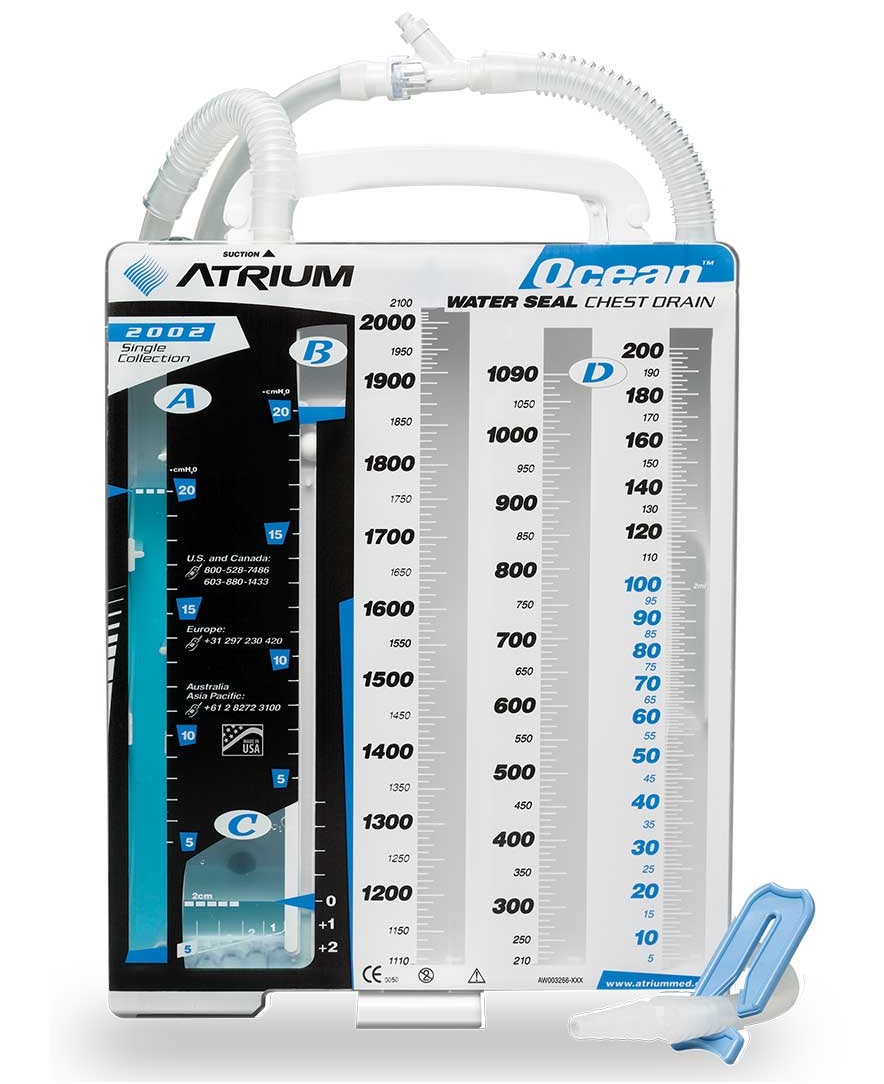
A patient with a chest tube requires the healthcare team to perform frequent assessments, including: Chest tubes also require additional care. The care of chest tubes involves:Nursing Assessments and Care
Ensure the water seal stays at 2 cm or what the provider ordered. The water seal is crucial to create the one-way valve. If the water seal is not present, the drainage system will not work.Water Seal
The provider will routinely evaluate the character of the output in the collection chamber. They will assess if the output is serosanguinous. It is normal to see a bit of serosanguinous output upon initial insertion of the chest tube. However, a lot of serosanguinous output can also indicate hemorrhage.Character of Output
The provider will routinely evaluate the amount of output. As a general rule, an output of over 100 mL per hour is cause for concern because it can place the patient at risk for hypovolemia and associated complications.Amount of Output
Related Video – One Quick Question: How Do You Calculate Tube Depth?
The provider will routinely check for bubbling in the water seal chamber. Continuous bubbling is concerning because it can indicate a leak. However, intermittent bubbling is normal and occurs when the patient coughs or exhales. Intermittent bubbling can also occur when a patient has a pneumothorax because air is leaving the pleural space. That is also normal.Bubbling
The provider will observe that there is tidaling of the water seal chamber. This means that the water seal chamber rises with inspiration and falls with expiration. It is normal to see fluctuation in the water seal chamber, as it indicates the chest tube connection with the chest is optimal.Tidaling
The provider ensures that the chest tube and collection chamber are always kept below the patient’s chest level, otherwise the contents in the collection chamber will flow back into the patient.Relative Location to the Chest
Milking the tube occurs when the tube is squeezed to remove obstructions. Milking the tube can potentially cause tension pneumothorax. However, if a clot is observed in the tube, it is okay for the provider to gently milk the tube.Avoid Milking the Tube
If the chest tube comes out of the patient, the incision hole needs to be covered with an occlusive material. Therefore, petroleum gauze should be kept by the patient’s bedside. When applied, the gauze is taped on three of the four sides. Taping the gauze on all four sides can potentially plug up the hole and cause tension pneumothorax. Air needs to be allowed to exit but not reenter the hole. When the patient breathes in, the patch sticks to the skin to keep air from going back into the chest cavity.Petroleum Gauze and Tape
Sterile water is used to ensure the water seal chamber is at the appropriate level. Also, in case the chest tube disconnects from the collection chamber, the chest tube should be placed in sterile water to maintain the one-way valve until the tube can be reconnected.Sterile Water
Every chest tube includes a clamp. The provider will determine if and when the tube should be clamped. Clamping the tube has the potential to cause tension pneumothorax. The clamp is only used under certain circumstances, such as changing the chest drainage unit. A chest tube drains liquid from a pleural effusion. Chest tubes are used in certain circumstances to facilitate lung expansion or to remove fluid from the chest cavity. They require special attention and care from the healthcare team.Clamping
More Free Resources to Keep You at Your Best
Editorial Note
ACLS Certification Association (ACA) uses only high-quality medical resources and peer-reviewed studies to support the facts within our articles. Explore our editorial process to learn how our content reflects clinical accuracy and the latest best practices in medicine. As an ACA Authorized Training Center, all content is reviewed for medical accuracy by the ACA Medical Review Board.
